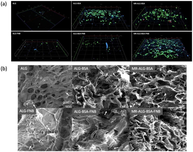
Figure 5.
AREP-19 cells seeded on ALG-based scaffolds. (a) 3D reconstruction of confocal microscope images of seeded ARPE-19 cells on scaffolds with fluorescence staining after 14 days: DAPI (blue) calcein-AM (green) PI (red) in the depth of 200 µm of scaffolds (scale bar: 100 µm).; y = 1200 µm, x = 1200 µm, thickness = 200 µm, (b) SEM images of cells distributed on the ALG-based scaffolds; (Scale bar: 100 µm). White arrows indicate the presence of ARPRE-19 cells.

