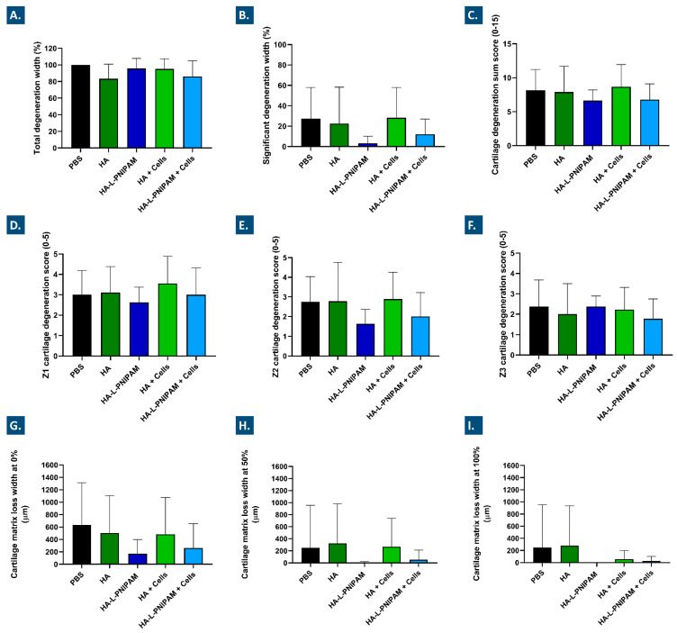
Figure 6.
Medial tibial plateau histomorphometry evaluation of total (A) and significant (B) cartilage degeneration width. A percentage relative to the total width of the articular surface was used for total and significant cartilage degeneration width evaluation. A cartilage degeneration sum score for the medial tibial plateau was obtained by combining the scores of the three plateau sections (i.e., Z1 + Z2 + Z3) (C), considering the external third of the plateau (i.e., Z1) (D), the central third of the plateau (i.e., Z2) (E), and the internal third of the plateau (i.e., Z3) (F). Cartilage matrix loss width values were measured in the medial tibial plateau at 0% (i.e., at the surface, (G)), at 50% cartilage depth (H), and at 100% (I) cartilage depth, respectively. All results are expressed as means with standard deviations as error bars for eight or nine animals per group. HA, hyaluronic acid; PNIPAM, poly(N-isopropylacrylamide).

