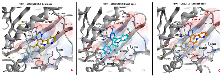
Figure 7.
Representation of the selected pose produced by molecular docking with the program PLANTS for the compound 5d (colored in gold, panel A), 5j (depicted in cyan, panel B), and 4e (colored in orange, panel C) in the ATP-binding site of the ligand-based homology model created for PDK1. All the selected conformations passed the steric and electrostatic filtering processes. In each panel, also the electrostatic surface around the ligand in the binding site is represented. The images were created and rendered with MOE.

