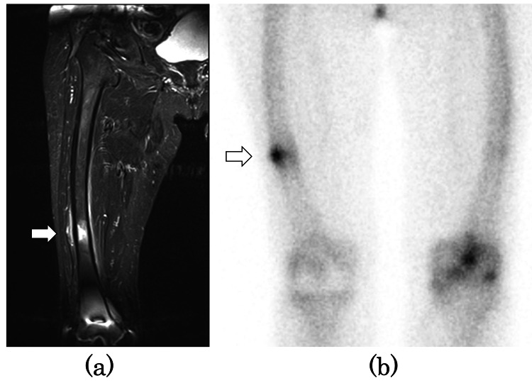
Figure 2. Preoperative MRI STIR coronal image and bone scintigraphy.
(a) MRI STIR coronal image showed bone marrow edema in the diaphysis of the right femur (white arrow). (b) Bone scintigraphy showed a stress fracture of the right femur (white arrow).
MRI: magnetic resonance imaging, STIR: short tau inversion recovery

