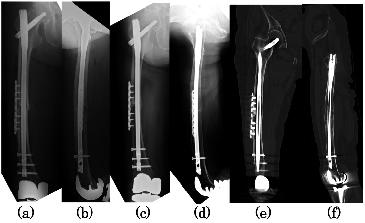
Figure 4. Radiographs taken immediately after surgery and at one year and two months postoperatively.
Immediate postoperative radiographs: (a) anteroposterior and (b) lateral images. Radiographs at one year and two months postoperatively: (c) anteroposterior and (d) lateral images. Plain computed tomography at one year and two months postoperatively: (e) coronal and (f) sagittal images. Anterior, posterior, and medial bony fusion of the osteotomy has been achieved, and bony bridging has been achieved on the lateral side.

