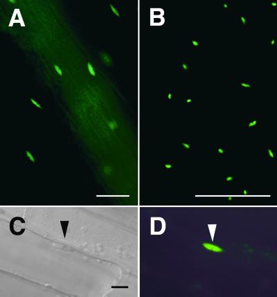
Figure 3.
Nuclear Localization of GFP–CIP4.
Transgenic Arabidopsis expressing GFP–CIP4 was examined under a light microscope.
(A) Fluorescence microscope image of a hypocotyl.
(B) Fluorescence microscope image of a leaf.
(C) Differential interference contrast microscope image of hypocotyl cells.
(D) Florescence microscope image of the same hypocotyl cells shown in (C).
The large track of diffused fluorescence signal from top left to bottom right in (A) is due to a vascular bundle that was out of focus. The positions of GFP fluorescence were confirmed to be nuclei by differential interference contrast imaging or 4′,6-diamidino-2-phenylindole staining (data not shown). The triangles in (C) and (D) point to the nucleus.  ;
;  for (C) and (D).

