
Figure 1.
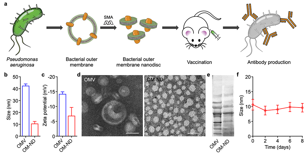
Preparation and characterization of OM-NDs. (a) Bacterial OMVs are collected from Pseudomonas aeruginosa and incubated with styrene-maleic acid (SMA). The resulting OM-ND formulation can be used as a nanovaccine to protect against bacterial infection. (b,c) Size (b) and zeta potential (c) of free OMVs and OM-NDs (n = 3, mean + SD). (d) Transmission electron microscopy images of free OMVs (left) and OM-NDs (right) after negative staining (scale bars: 20 nm). (e) Protein profile of OMVs and OM-NDs after gel electrophoresis. (f) Size of OM-NDs in PBS over time (n = 3, mean ± SD).