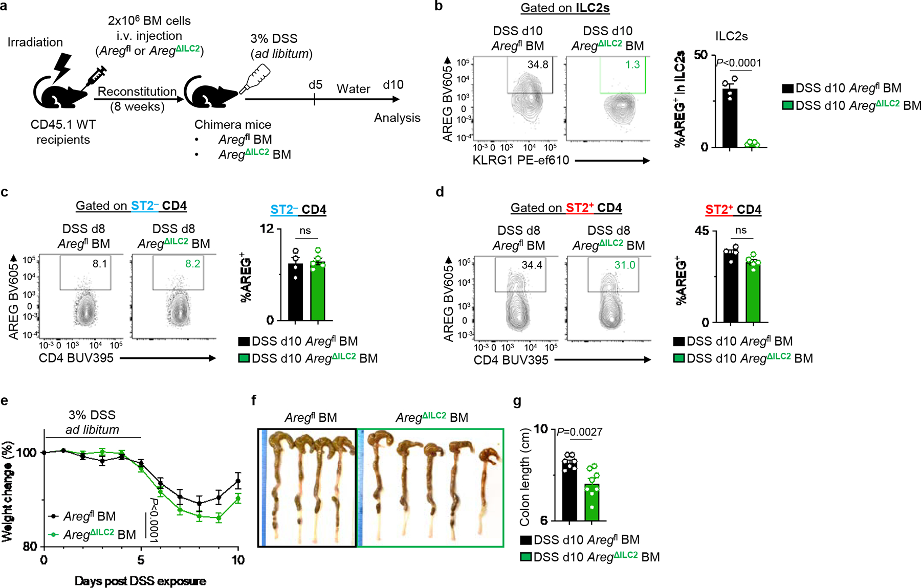
Extended Data Figure 8 |. Bone marrow chimera mice recapitulated the phenotype of AregΔILC2 mice in the context of DSS-induced intestinal damage and inflammation.

Irradiated CD45.1 WT mice received BM from Aregfl or AregΔILC2 donor mice. After 8 weeks, chimera mice were exposed to 3% DSS for 5 days and then allowed to recover for 5 days on regular drinking water. a, Experimental schematic. b-d, Examination of AREG deletion in ILC2 (b), ST2− CD4+ T cells (c), and ST2+ CD4+ T cells (d) in DSS-exposed Aregfl BM (n=4 mice) and AregΔILC2 BM (n=5 mice) by flow cytometry. e-g, Disease severity of DSS-exposed Aregfl BM (n=7 mice) or AregΔILC2 BM (n=8 mice) as determined by weight loss (e) and colon length (f, g). Data in b-d are representative of three independent experiments. Data in e and g are pooled from 2 independent experiments. Unpaired two-sided t-test (b-d and g). Two-way ANOVA with Šídák multiple comparisons test (e). P values are presented where appropriate. ns, not significant. Data are represented as means ± S.E.M.
