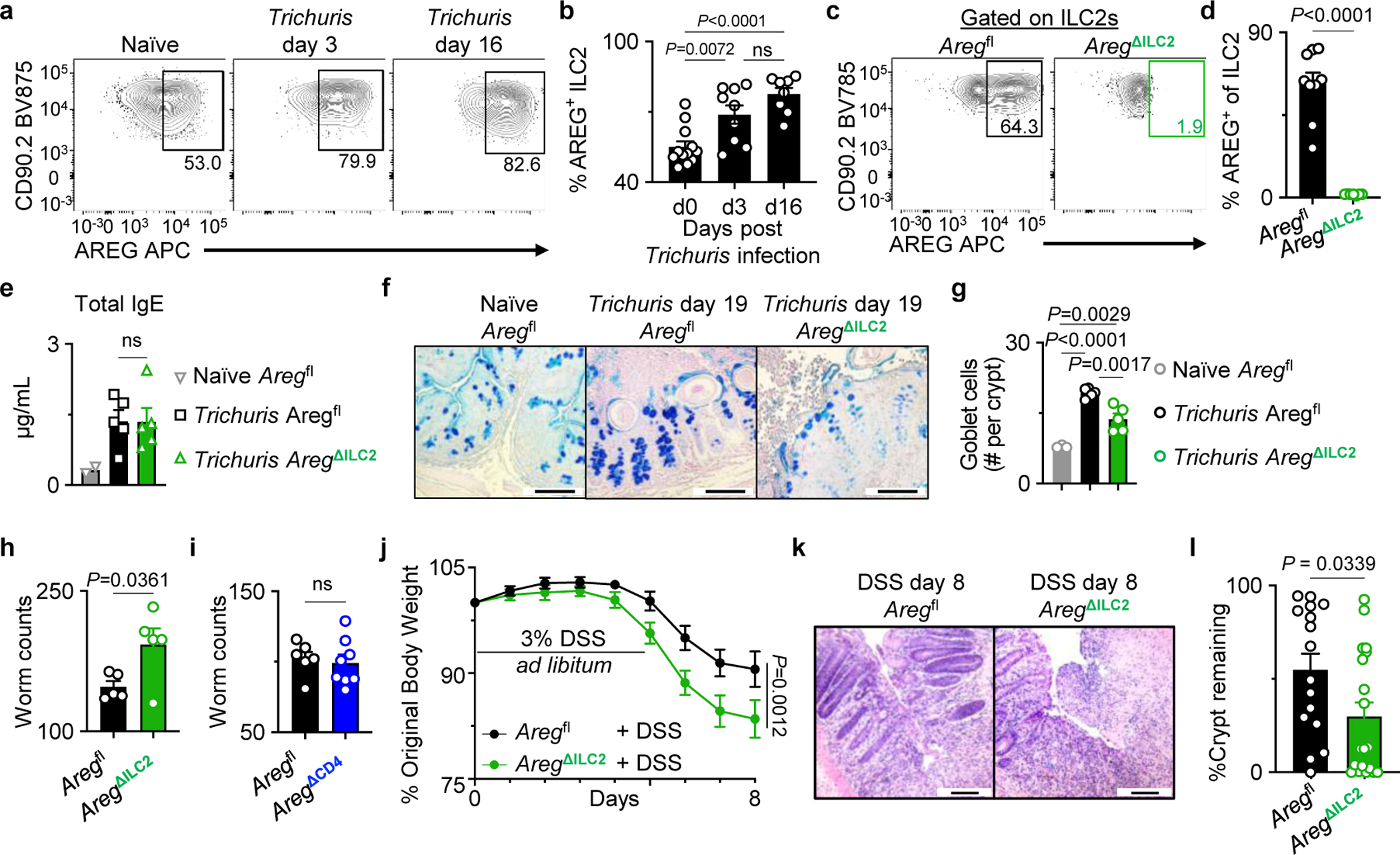
Figure 3 |. ILC2-derived AREG is critical for clearance of the gut-dwelling helminth Trichuris muris and tissue protection following intestinal injury.

a, b, Representative flow cytometry analysis of ILC2s (a) and percentages of AREG+ ILC2s isolated from WT mice at day 0 (n=12 mice), day 3 (n=9 mice), and day 16 (n=8 mice) post-Trichuris infection (b). c, d, Representative flow cytometry analysis of ILC2s (c) and percentages of AREG+ ILC2s in the colons of Aregfl (n=9 mice) or AregΔILC2 (n=7 mice) (d). e, Total serum IgE levels from naïve Aregfl (n=2 mice) and Trichuris-infected Aregfl (n=5 mice) or AregΔILC2 (n=5 mice) analyzed at day 19 post-infection. f, g, Goblet cells in the proximal colon of naïve Aregfl (n=3 mice) and Trichuris-infected Aregfl (n=5 mice) or AregΔILC2 (n=5 mice) analyzed at day 19 post-infection by AB-PAS staining (bars=100 μm) (f). Enumerated goblet cells per crypt (g). h, i, Trichuris worm counts from Aregfl (n=5 mice) and AregΔILC2 (n=5 mice) (h) or Aregfl (n=6 mice) and AregΔCD4 (n=8 mice) (i) on day 19 post infection. j-l, Measurements of disease severity of DSS-exposed Aregfl (n=16 mice) and AregΔILC2 (n=19 mice) as determined by weight loss (j) and colon epithelial crypt architecture analyzed by H&E staining (bars=100 μm) (k, l). Data in b and d are pooled from two independent experiments. Data in e, g, h are representative of three independent experiments. Data in i are representative of two independent experiments. Data in j and l are pooled from four independent experiments. One-way ANOVA with Tukey multiple comparisons test (b, e, g). Unpaired two-sided t-test (d, h, i, l). Two-way ANOVA with Šídák multiple comparisons test (j). P values are presented where appropriate. ns, not significant. Data are represented as means ± S.E.M.
