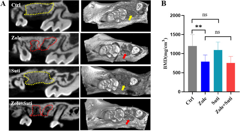
Fig. 2.
Interference of new alveolar bone formation and alveolar bone remodeling activity in extraction sockets after application of anti-resorptive drugs. A Coronal section and 3D reconstruction of micro-CT images of the maxillary bone after two weeks after the left maxillary second molar extraction. The positions pointed by the yellow dotted line and arrow indicate that the new formed alveolar bone of the extracted tooth is well remodeled, while the red dotted lines and arrows is labeled that the socket bone of the extracted tooth is poorly remodeled. B Statistical analysis of bone mineral density of newly formed alveolar bone in extraction socket. BMD, bone mineral density

