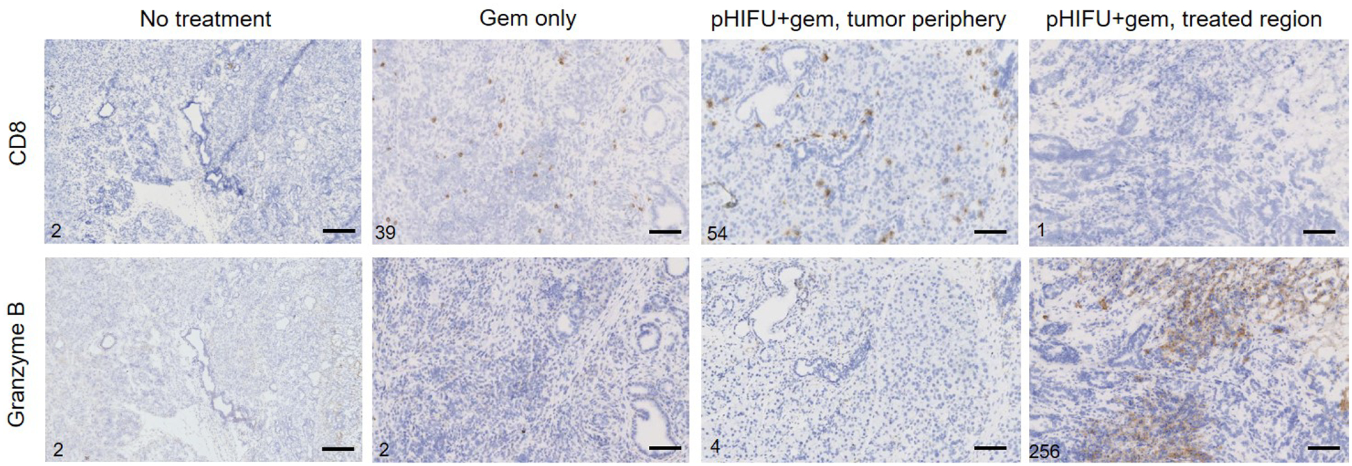
Figure 4.

Serial CD8 and Granzyme B stained sections of tumors from the experimental groups. The numbers of positive cells seen within each frame are provided in the lower left corner of the frame. CD8-positive cells were present in similar numbers in well differentiated, peripheral areas of tumors from both treatment groups, and lower numbers in control group, but with little to no Granzyme-B-positive staining, and were absent from less differentiated areas, the tumor core and pHIFU-treated regions. Conversely, pHIFU-treated regions consistently had intense Granzyme-B-positive staining, with a mixture of diffuse and focal staining that could indicate the presence and activation of immune cells other than CD8+ T cells in those areas. The scale bar in all images is 250 μm.
