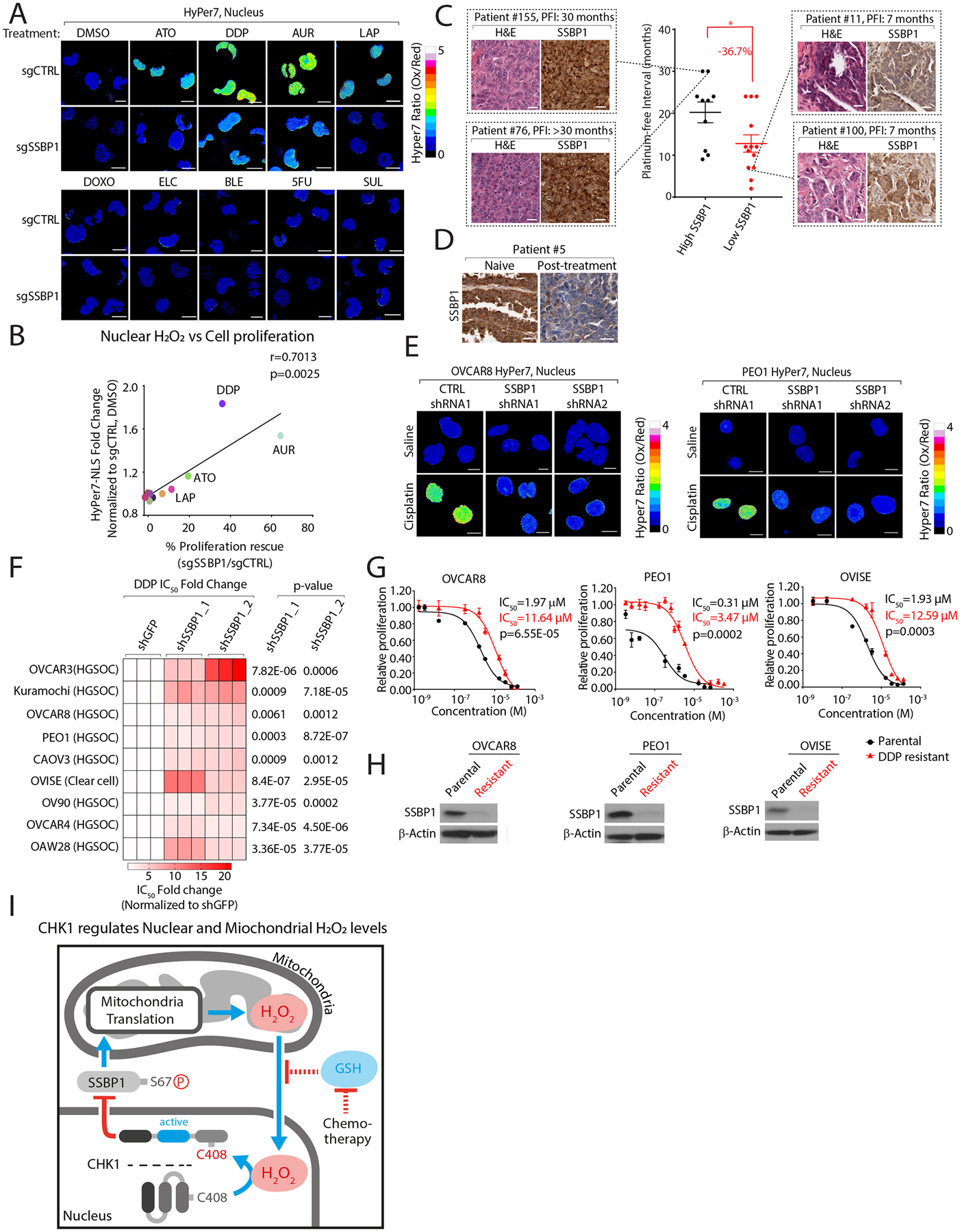
Figure 7: SSBP1 regulates nuclear H2O2 levels and mediates cisplatin resistance in ovarian cancer cells.

(A) SSBP1 depletion decreases nuclear H2O2 levels following treatment with anticancer agents in K562 cells. (B) Comparison of fold-change in nuclear H2O2 levels with proliferation rescue in K562 cells depleted of SSBP1 following treatment with the indicated compounds. (C) Lower SSBP1 levels correlate with shorter platinum free intervals (PFI) in high-grade serous ovarian cancer (HGSOC) tumors. (D) SSPB1 levels are decreased in platinum-refractory HGSOC tumors. (E) Knockdown of SSBP1 decreases cisplatin regulated nuclear H2O2 levels in ovarian cancer cell lines. (F) Heatmap depicting fold change in DDP IC50 values in ovarian cancer cell lines expressing the indicated shRNAs targeting SSBP1. (G-H) DDP-resistant ovarian cancers have decreased SSBP1 expression. (G) DDP IC50 values were measured in parental or DDP-resistant ovarian cancer cell lines. (H) Immunoblot analysis of SSBP1 in the indicated cell lines. (I) Model. Nuclear H2O2 activates CHK1 leading to the phosphorylation and cytosolic retention of SSBP1. Cytosolic SSBP1 cannot promote mitochondrial translation which generates H2O2. Mitochondrial H2O2 is transmitted to the nucleus following a decrease in GSH:GSSH ratio by certain anticancer drugs. Data are represented as mean ± SEM. * p < 0.05, ***p< 0.0001. Statistical significance was determined by one-way ANOVA with Sidak’s post-hoc correction.
