Figure 2.
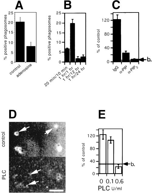
Involvement of PI(4,5)P2 in actin assembly. (A) Actin assembly on 2-h phagosomes in the absence (control) or presence of 200 μM adenosine. (B) Phagosomes purified after different pulse/chase times of beads in cells were analyzed for their PI(4,5)P2 content, via antibody labeling, followed by flow cytometry analysis. The same treatment of noninternalized beads, Triton X-100–pretreated phagosomes, or incubation of phagosomes with the secondary antibody alone gave no detectable signal (data not shown). (C) 2-h phagosomes were preincubated for 5 min at 25°C with an irrelevant antibody (IgG), anti-PI(4)P antibodies (α-PIP), or anti-PI(4,5)P2 antibodies (α-PIP2). Actin assembly on phagosomes after antibody preincubation was assayed by microscopy. Results show the percentage of positive phagosomes for each sample relative to control phagosomes. (D) 2-h phagosomes were pretreated with 0 (control) or 0.6 U/ml bacterial PI-PLC and immediately labeled for PI(4,5)P2 by indirect immunofluorescence microscopy. Arrows show individual phagosomes seen by phase-contrast microscopy. Bar, 5 μm. (E) In parallel experiments, 2-h phagosomes were pretreated in the absence or presence of 0.1 or 0.6 U/ml bacterial PI-PLC. Actin assembly on subsequently reisolated phagosomes was then assayed by microscopy. RESULTS show the percentage of positive phagosomes for each sample relative to control (untreated) phagosomes. b indicates the value obtained with fish-skin gelatin–coated beads.

