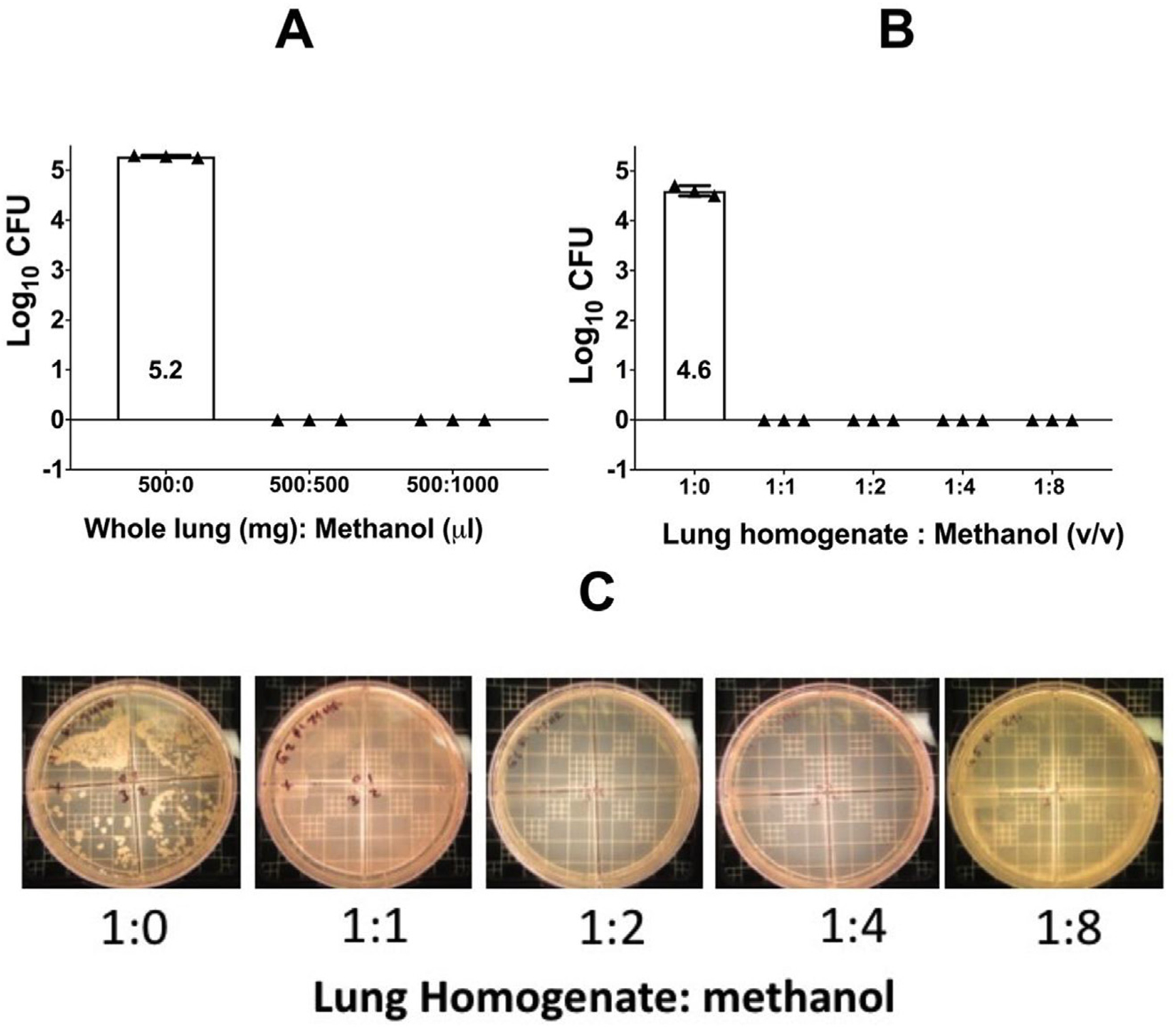
Fig. 1. CFU growth of lung specimens after methanol sterilization.

Panel A shows CFU growth of untreated whole lung compared to 1:1 and 1:2 tissue to methanol (mg/ul). Panel B shows CFU growth of untreated lung homogenate compared to 1:1 and 1:2 tissue to methanol (v/v). Panel C shows CFU growth of lung homogenate on 7H11 agar after 6 weeks incubation at 37 °C and diluted ten-fold. Homogenate was treated with 0 μl of methanol or 1:1, 1:2, 1:4, or 1:8 (v/v) of methanol as shown.
