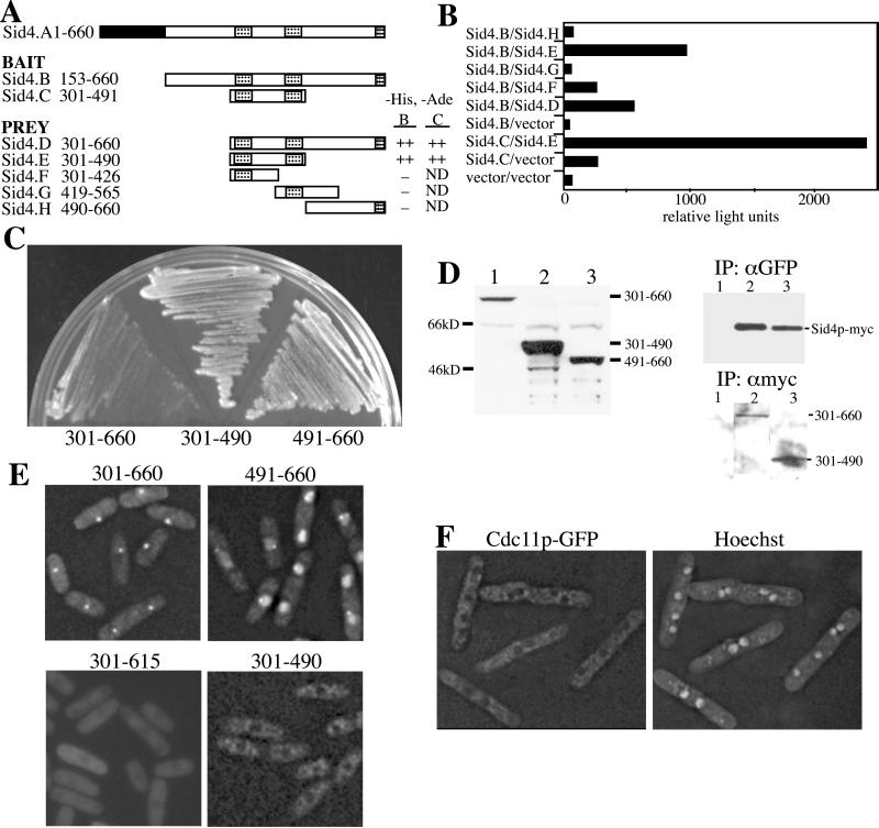
Figure 6.
The Sid4p C terminus binds SPBs and displaces Cdc11p. The indicated regions of Sid4p in either bait or prey plasmids were cotransformed into S. cerevisiae strain PJ69–4A. Leu+Trp+ transformants with either bait “B” or “C” fragments were scored for growth on selective medium (A) or for β-galactosidase activity (B) that was measured in relative light units. The three coiled-coil domains of Sid4p are indicated in the diagram by hatched boxes. (C) Wild-type cells (KGY246) expressing the indicated GFP-Sid4p fusion proteins from the full-strength nmt1 promoter were struck to medium lacking thiamine at 32°C. Colonies were allowed to form for 3 days. (D and E) The sid4-myc (KGY1340) (D) and wild-type (KGY246) (E) strains were transformed with pREP81 vectors directing production of the indicated GFP-Sid4p fusion proteins. Transformants were grown in the absence of thiamine and either protein lysates were prepared (D) or live cell images captured (E). A sample of each lysate was resolved by SDS-PAGE and immunoblotted with antibodies to GFP (D, left panel). The remaining lysates were divided in half and subjected to immunoprecipitation with either polyclonal anti-GFP serum or the 9E10 monoclonal anti-myc antibody. Immunoprecipitates were resolved by SDS-PAGE and immunoblotted with 9E10 (D, right upper panel) or anti-GFP serum (right lower panel). (F) The cdc11-GFP strain (KGY3341) was transformed with pREP1sid4ΔNp (residues 153–660) and grown in the absence of thiamine for 20 h. Images of live cells stained with Hoechst were captured.

