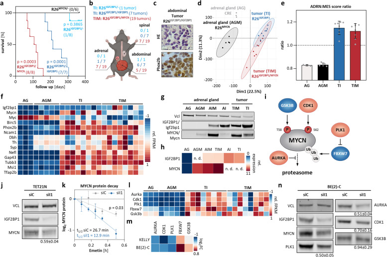
Fig. 6.
IGF2BP1 induces neuroblastoma, Mycn expression, stabilizes MYCN protein and synergizes with MYCN in transgenic mice. a Kaplan–Meier survival analysis of heterozygous (cyan, n = 8) and homozygous (blue, n = 7) R26IGF2BP1, heterozygous R26MYCN (black, n = 6) and double transgenic R26IGF2BP1/MYCN (red, n = 8) mice. Numbers in brackets indicate tumor bearing mice. b Scheme of tumor location within mice. c Representative images of hematoxylin and eosin staining (HE, top) and Phox2b immunohistochemistry (bottom) indicative for neuroblastoma in R26IGF2BP1/IGF2BP1 mice (bars: 40 µm). d PCA of mouse adrenal glands and transgenic tumors. e Ratio of ADRN to MES signature of mouse adrenal glands and transgenic tumors. f Heatmap depicting row-scaled FPKM values of indicated murine mRNAs in adrenal glands derived from wildtype (n = 3, AG) or R26MYCN/− (n = 5, AGM) mice and tumors of R26IGF2BP1 (n = 8, TI) or R26IGF2BP1/MYCN (n = 8, TIM) mice. g Western blot analysis confirms IGF2BP1 or MYCN transgene expression in tumors and adrenal glands of representative mice (n = 1). h RT-qPCR analysis of human IGF2BP1 and MYCN mRNA in indicated mouse tissue (n. d.—not determined). i Scheme of MYCN protein regulation. j Western blot (n = 5) analysis of MYCN expression upon IGF2BP1 (siI1) compared to control (siC) knockdown in TET21N cells. k MYCN protein decay was monitored by Western Blot analysis in control (grey) and IGF2BP1 knockdown (blue) TET21N cells upon indicated time of emetin treatment (n = 3). l, m RNA-seq analysis of indicated mRNAs in mouse tissues (l) or upon transient IGF2BP1 knockdown in BE(2)-C and KELLY (m). n Western blot (n = 3) analysis of indicated proteins upon IGF2BP1 (siI1) compared to control (siC) knockdown in BE(2)-C

