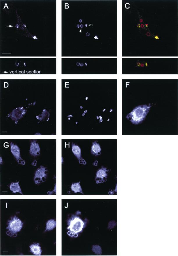
Figure 3.
Early FcγR-mediated signaling and recruitment events proceed normally in macrophages expressing dominant negative Vav. (A–F) FcγR-mediated phagocytosis of RBC is accompanied by the underlying accumulation of phosphotyrosine proteins in control (A–C) and in Vav-C expressing (D–F) macrophages. Endogenous phosphotyrosine proteins (A), RBC (B), and merged image (C) showing RBC in red and endogenous tyrosine-phosphorylated proteins in green. Note the transient nature of the local enrichment in phosphotyrosine staining; in B, open arrowhead indicates bound RBC (phosphotyrosine positive), whereas closed arrowhead shows fully internalized RBC (negative for phosphotyrosine proteins). Expression of Vav-C (F) fails to alter the enrichment of phosphotyrosine proteins (D) beneath bound RBC (E). (G–J) Recruitment of the protein tyrosine kinase Syk, which accompanies FcγR-mediated engulfment in macrophages, proceeds normally in control (G–H) and Vav-C–expressing cells (I–J). Endogenous Syk (G and I), IgG-opsonized latex beads (H), and Vav-C and IgG-opsonized latex beads (both detected in J, due to cross-reactivity of the secondary antibody). Latex beads were used to overcome difficulties in staining for Syk. Representative images from four independent experiments are shown. Bar, 10 μm.

