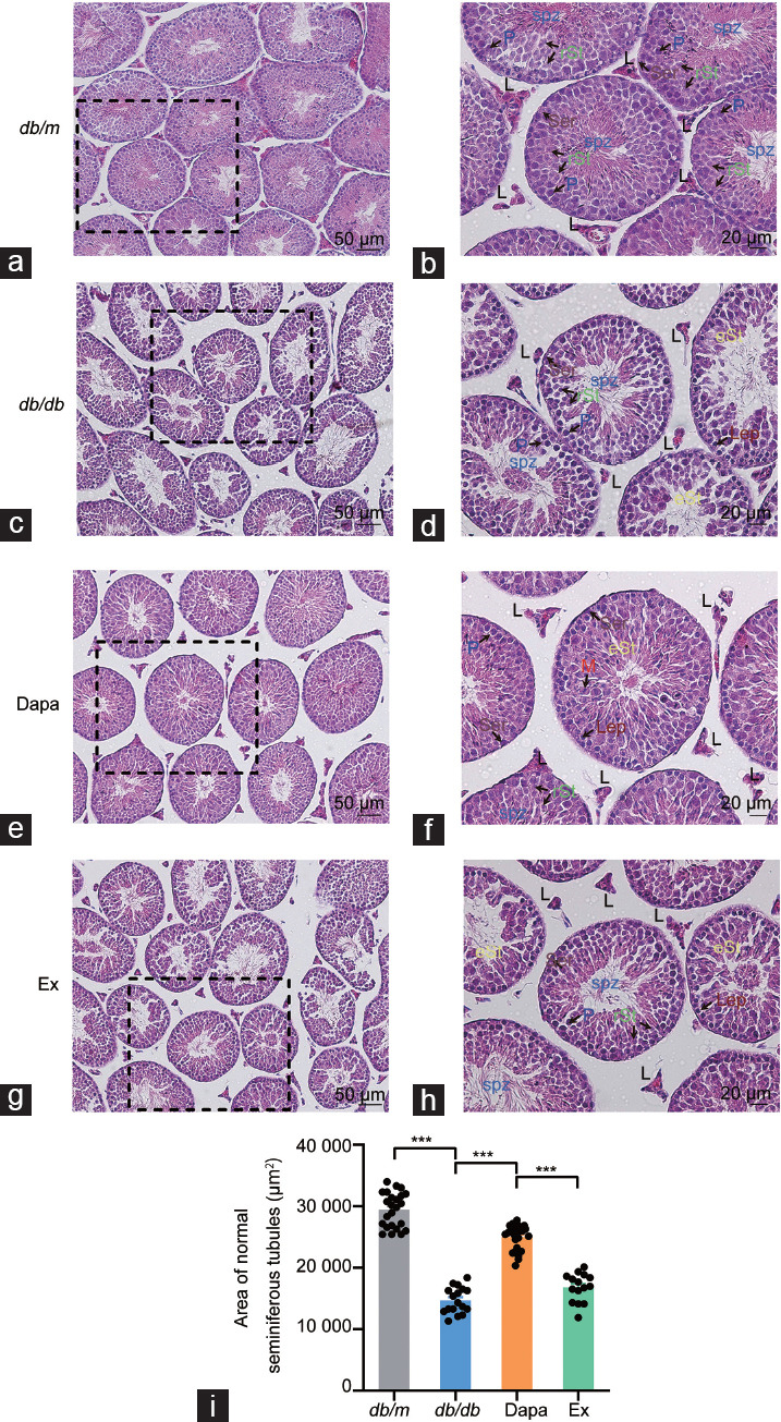
Figure 1.

H&E staining of the testicular tissue of db/m mice and db/db mice treated with dapagliflozin (Dapa) and dapagliflozin + exendin (9–39) (Ex). H&E staining of the testicular tissue of db/m mice with (a) scale bar = 50 µm and (b) scale bar = 20 µm. H&E staining of the testicular tissue of db/db mice with (c) scale bar = 50 µm and (d) scale bar = 20 µm. H&E staining of the testicular tissue of Dapa mice with (e) scale bar = 50 µm and (f) scale bar = 20 µm. H&E staining of the testicular tissue of Ex mice with (g) scale bar = 50 µm and (h) scale bar = 20 µm. Arrows with abbreviations in different colors indicate different types of cells. (i) Area of normal seminiferous tubules. n = 4 mice per group. All data are presented as the mean ± standard error of mean. ***P < 0.001. H&E: hematoxylin and eosin; P: pachytene spermatocytes; rSt: round spermatids; eSt: elongating spermatids; Lep: leptotene spermatocytes; M: meiotic spermatocytes; L: Leydig cells; Ser: Sertoli cells.
