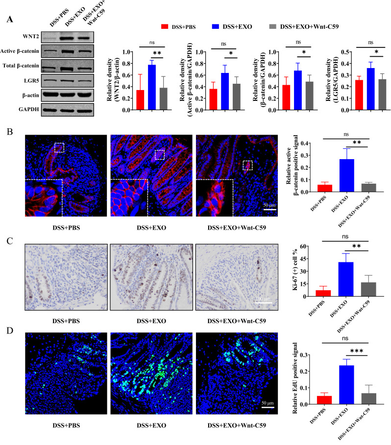
Figure 7.
Wnt-C59 blocked the promoting effects of hucMSC-exosomes on regeneration of ISCs and intestinal epithelium partially. (A) Western blot analysis of the protein expression level of WNT2, active β-catenin, total β-catenin and LGR5 in the pathway inhibited model (n=5, corresponding full-length blots are presented in Supplementary Figure 4). (B) The localization and expression of active β-catenin (red) was analyzed by IF staining of active β-catenin. The purple region produced by the mixture of red and blue (DAPI) was thought to be the place where β-catenin entered the nucleus. The proliferation potential of colon epithelium was measured by IHC staining of Ki-67 ((C) n=5, scale bar=100 µm, 400×) and EdU incorporation assay ((D) n=5, scale bar=50 µm, 400×). Data were shown as mean ± SD. The Mann–Whitney U-test (non-normal distribution) or Unpaired Student’s t-test (normal distribution) was used to compare the variables between two groups. P<0.05 was considered as statistically significant. *P<0.05, **P<0.01, ***P<0.001, ns indicates P>0.05.

