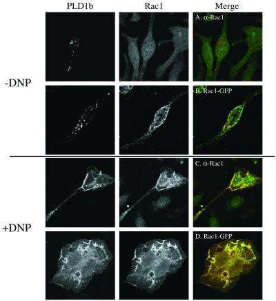
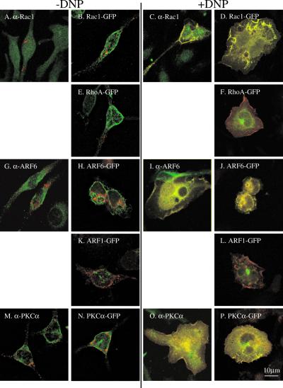
Figure 2.
Antigen-stimulated colocalization of PLD1b with Rac1, ARF6, and PKCα. The FcεRIs of RBL-2H3 cells were sensitized by the addition of 1 μg/ml anti-DNP IgE for 16 h. Confocal images were obtained from cells before receptor cross-linking with DNP (−DNP; A, B, E, G, H, K, M, and N) or after addition of 50 ng/ml DNP-HSA for up to 8 min (+DNP; C, D, F, I, J, L, O, and P). Transfected PLD1b is indicated by the red staining. Transfected or endogenous, antibody-detected regulators of PLD1b are indicated by the green staining. Colocalization of PLD1b and its regulator is indicated by yellow staining. The first part of this figure shows the Rac1 experiments and is used as an example of the staining for individual proteins. In the rest of this figure only the merged images are shown. Identical results were obtained with PLD1b HA- or GFP-tagged and Rac1- and ARF6 both tagged with either HA- or GFP (our unpublished results). Similar results were obtained in at least three experiments.


