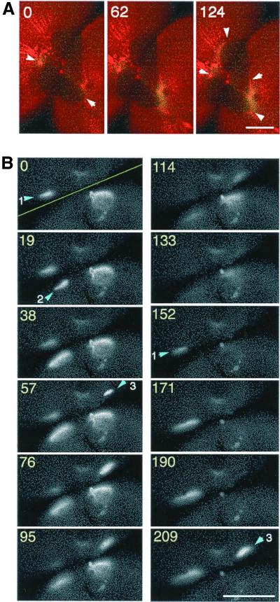
Figure 6.
Ca wave 2 propagates along the border of old and new membranes after completion of cytokinesis. (A) Representative merged image of rhodamine-WGA staining (red) and increasing free [Ca2+]i: (FCalG − FCNF) × 2 (green) during Ca wave 2 after completion of the first cleavage. Wave 2 emerged at the animal pole and traversed along the border of old and new membranes. Arrowheads in the image at 0 s indicate the starting point of wave 2, and those in the image at 124 s indicate the wave front. Note that the egg had already divided into two blastomeres before wave 2 first appeared. (B) Gray scale images of increased free [Ca2+]i; (FCalG − FRhod) during a typical wave 2. The yellow line in “time 0” indicates the position of the division plane. Wave 2 does not propagate continuously, but often skips a distance of several hundred micrometers, reappearing along the border of the two membranes or flickering at some particular site several times. Arrowheads indicate initiation sites of wave 2. At site 1, Ca signal is flickering (the signal is seen during 0–95 s, disappears and then reappears after 152 s). There seemed to be no synchrony between the waves in the two blastomeres. Numbers indicate the recording time (seconds). Scale bars, 0.2 mm.

