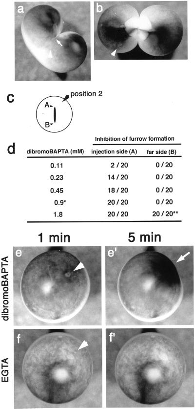
Figure 9.
Effect of dibromoBAPTA and EGTA on the cortex of Xenopus eggs. (a and b) Thirty minutes after the injection of 0.45 mM dibromoBAPTA (a) or injection buffer (b) into dividing wild-type Xenopus egg. Arrowheads, injection sites; white arrow, the position of the growing end. (a) Furrow formation was clearly inhibited on the side of the injection. The growing end was drawn back to the center of the egg after its progression had stopped. On the other hand, the furrow progressed normally at the other end. In the control egg (b), both ends grew and the egg cleaved normally. (c) Positions of injection sites. Position 2, injection carried out at a position near the growing end of the cleavage furrow. (d) Dose-dependent inhibition of furrow formation by dibromoBAPTA. Progression of the furrow at the growing ends (A or B) was examined 30 min after injection. ∗, concentration (0.9 mM) used in Ca monitoring experiment in Figure 8. ∗∗, at 1.8 mM, progression of the furrow at the end B was very slow, and the egg was severely deformed. Ca buffers were injected into fertilized wild-type Xenopus eggs, and changes of the cortex were monitored 1 min (e and f) and 5 min (e′ and f′) after the injections. Arrowheads in (e) and (f) indicate the positions of injection site. Injection of 1.8 mM dibromoBAPTA (e′), induced deformation of cortex and concentration of pigment granules around the injection site (arrow). (f and f′) Injection of 6.3 mM EGTA did not cause such contraction in the cortex.

