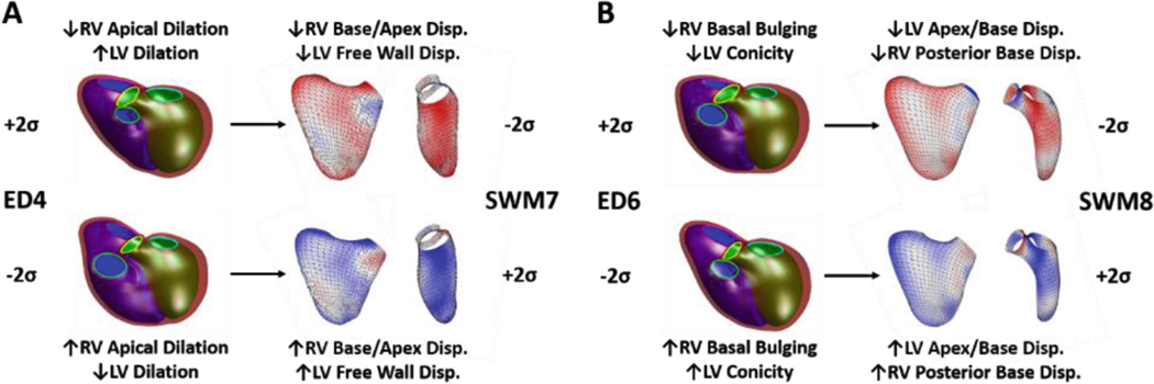
Fig. 2.
Summary morphological characteristics for associations between ED shape and SWM, (A) ED4 ∝ SWM7 and (B) ED6 ∝ SWM8. For ED shape modes, the LV endocardial surface, RV endocardial surface, and epicardial surface are shown in green, blue, and maroon, respectively, and the mitral, tricuspid, aortic, and pulmonary valves are shown in cyan, pink, yellow, and green, respectively. For SWM modes, the LV and RV free walls are shown and are colored based on the systolic displacement relative to the mean, inward (blue) and outward (red).

