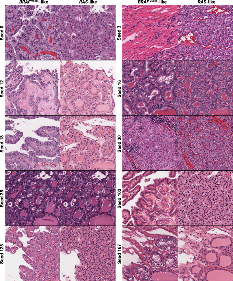
Fig. 3. cGAN-generated synthetic histology illustrates morphologic differences associated with BRAF-RAS gene expression score in thyroid neoplasms.
Classifier-concordant seeds from the thyroid cGAN were reviewed with an expert thyroid pathologist and pathology fellow to determine thematic differences in cGAN-generated BRAFV600E-like and RAS-like histologic features. Seed 0 illustrates architectural differences, with a papillae in the BRAFV600E-like image replaced with colloid in the RAS-like image. Seed 3 highlights the an increase in fibrosis in the BRAFV600E-like image. The BRAFV600E-like image for seed 12 shows a cystic structure with cell lining, which is replaced with what appears to be a tear in the RAS-like image, accompanied by architectural differences moving from papillae in the BRAFV600E-like image to follicles in the RAS-like image. Seed 16 demonstrates an increase in lacunae caused by resorbed colloid in the BRAFV600E-like image, compared with smaller, more regular follicles in the RAS-like image. Seed 18 shows papillae, a papillary vessel, and a cystic structure in the BRAFV600E-like image replaced with follicles, colloid, and an endothelial-lined vessel in the RAS-like image, respectively. Seed 30 highlights more tumor-infiltrating lymphocytes in the BRAFV600E-like along with increased cytoplasmic density compared with the RAS-like image. Seed 35 shows overall similar architecture in the two images, but with greater cell flattening in the RAS-like image compared to the BRAFV600E-like image. Seeds 102 and 128 both illustrate nuclear pleomorphism, increased cytoplasm, papillae, and scalloping in the BRAFV600E-like images compared with the RAS-like image. Seed 167 highlights nuclear pleomorphism and fibrosis in the BRAFV600E-like image compared with RAS-like image which has monotonous, circular, non-overlapping nuclei with regular contours and fine, dark chromatin.

