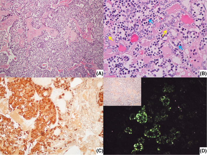
FIGURE 2.

(A) Low power view of mixed medullary‐papillary carcinoma on the right lobe (H & E, 100×); (B) Detail of the nuclei of the cells composing the tumor in A (small and round cells with clumped chromatin and finely granular cytoplasm—blue arrows—and cells with papillary features, that is, empty appearance of the nucleoplasm and nuclear irregularity and overlapping – yellow arrows) (H & E, 400×); C – Immunohistochemical staining for calcitonin highlights the medullary component (200×); D – Amyloid deposition, confirmed in polarized light, was already apparent using Congo red histochemistry (inset) (200×).
