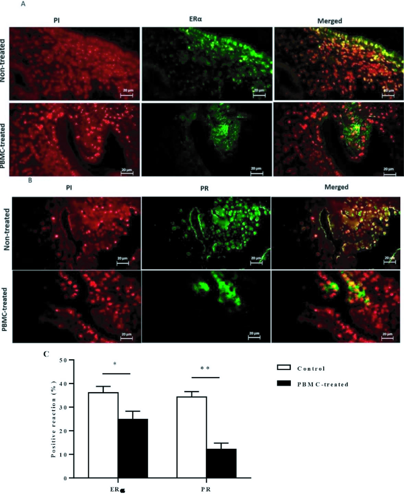
Figure 3.
A, B) ERα and PR protein expression in human endometrium. Fluorescent immunohistochemical analysis of ERα and PR protein expression in the endometrial section was carried out. Cell nuclei were stained with PI (red). Then ERα and PR specific signal and PI were merged, showing that ERα protein localization in the endometrial cells. C) The endometrial ERα and PR protein expression were significantly decreased in PBMC-treated (N = 10) compared to control group (N = 10). Endometrial tissue samples of each participant were evaluated in triplicate (*P 0.05, **P 0.01). Magnification = 400 (bar = 20 μm).

