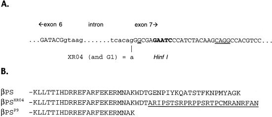
Figure 4.
(A) Nucleotide sequence around the boundaries of the cytoplasmic intron of myospheroid. Exon sequence in caps, intron in lowercase. The G>A mutation in mysXR04 (and mysG1) is indicated; this results in the seventh exon beginning with the underlined G. The potential downstream splice acceptor site is indicated by double underlining. To search for mRNAs that might use this site, this entire region was amplified by RT-PCR. Amplification of products using the conventional splice site was inhibited by cutting at the HinfI site indicated in bold (see text). (B) Predicted amino acid sequence of the cytoplasmic domains of wild-type βPS and that from mysXR04 and mysP9. The frameshift induced by the aberrant splicing in mysXR04 replaces the membrane-distal 21 residues with 25 essentially random amino acids (underlined). The first cytosine of exon 7 is a thymidine in mysG1; this polymorphism is silent in the parental strain, but changes to first residue encoded by the mutant exon 7 from alanine to valine.

