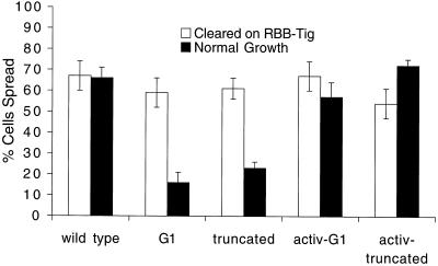
Figure 8.
Cell spreading in different assay conditions. Integrin induction in transformed S2 cells followed protease treatment as before (open bars) or was in the absence of protease (filled bars). In the latter case, the spreading assay was in normal growth medium, with serum, on uncoated tissue culture plates. activ-G1 and activ-truncation indicate cells that also express a mutant αPS2 that promotes integrin activation. The mutant β subunits support spreading poorly except in conditions (cleared cells or activating αPS2 subunits) that appear to promote artificially high activation. This is true in spite of the fact that the normal growth G1 and truncated cells express integrin at higher levels than any of the cleared or activating αPS2 lines.

