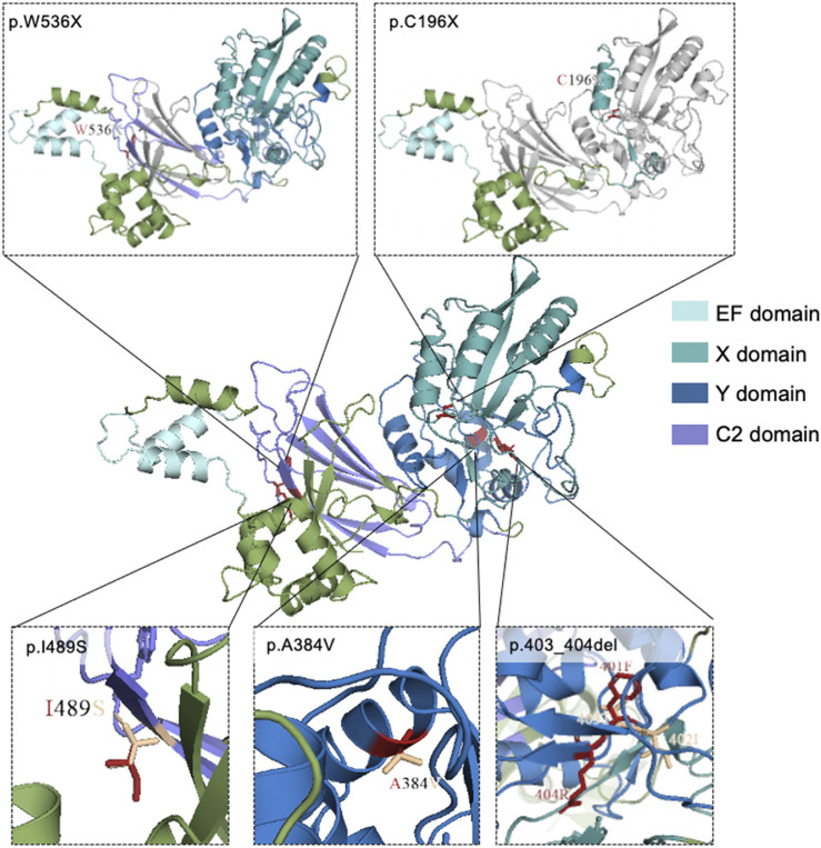
FIGURE 2.
Mutational locations in PLCZ1 proteins. 3D structure of wild-type and mutant models of PLCZ1 protein. Wild type protein structure was shown in the center. The above pictures show the truncated peptides of p.W536X and p.C196X respectively. Grey regions indicate the lost C-terminal after the new stop codon. The red residuals in the three pictures below show the residues of missense mutations, and the wheat residuals show the residues of the wild type amino acids.

