Figure 1.
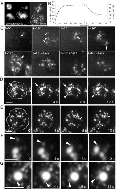
Endocytosis in living D. discoideum cells. (A) Comparison of epifluorescence microscopy and TIRM. Living D. discoideum cells were allowed to ingest rhodamine-green labeled dextran for 2 h, extracellular dextran was washed away to improve contrast, and endocytic compartments were visualized with both microscopes. (B) Biochemical measurements of kinetics of fluid phase uptake and egestion in wild-type D. discoideum cells (see MATERIALS AND METHODS). (C) Time course of fluid phase uptake and transit in wild-type D. discoideum cells. Cells were allowed to ingest rhodamine-green labeled dextran for different times (t = 5′ to 120′), briefly washed, and visualized by TIRM. To follow egestion, cells were fed with rhodamine-green dextran for 2 h, before incubation in dextran-free medium for the indicated times (t = 15′ chase to 60′ chase), and visualized by TIRM. Arrowheads point to dim early endosomes, arrows to small tubulo-vesicular endo-lysosomes, and asterisks highlight brighter late endosomal structures. (D–G) Endosomal morphology were studied in wild-type cells fed with rhodamine-green dextran for 3 h to ensure complete filling of all endocytic compartments and briefly washed with buffer; time-lapse series were recorded with TIRM. The cell boundaries are outlined in the first frame. (D) A big bright vacuole that was barely moving (asterisk), whereas small vesicles rapidly moved back and forth (arrow); a small group of vesicles and vacuoles that were very plastic and continuously changed shape are marked by arrowheads. The accompanying Movie 1 shows a longer time course (242 s) of the same cell. (E) Two very static vacuoles (asterisks) next to a very dynamic vacuole (dot) that continuously deformed and extended tubular structures; similar tubules extending from other vacuoles are marked by arrowheads. The accompanying Movie 2 shows a longer time course (246 s) of the same cell. Small vesicle fusing with a bigger vacuole (F); a bigger vacuole giving rise to a small vesicle (G). The movies play at 6 fps. Bars, 10 μm (C and D) and 5 μm (F and G).

