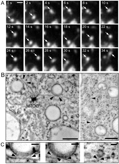
Figure 4.
Motility of endocytic vesicles. Wild-type cells were fed with rhodamine-green dextran for 3 h to ensure complete filling of all endocytic compartments and briefly washed with buffer; time-lapse series were recorded with TIRM. (A) A small bright vesicle undergoing rapid saltatory movements is marked by an arrow. The accompanying Movie 3 shows a longer time course (242 s) of the same cell. The movie plays at 6 fps; bars, 2 μm. (B) Ultrastructure of rapidly frozen wild-type D. discoideum cells. Vacuoles (470 and 390 nm in diameter) and small vesicles (120–130 nm) bound to microtubules by fine tethers are marked by arrowhead. The distance between microtubules and their bound vacuoles was 13 and 21 nm, respectively. The distance between microtubules and the smaller vesicles was significantly larger (30–40 nm). (C) Higher magnification of the tethers (arrowheads) that connect vacuoles and vesicles to the microtubules. MTOC, Microtubules organizing center. Bars, 0.5 μm (B) and 0.1 μm (C).

