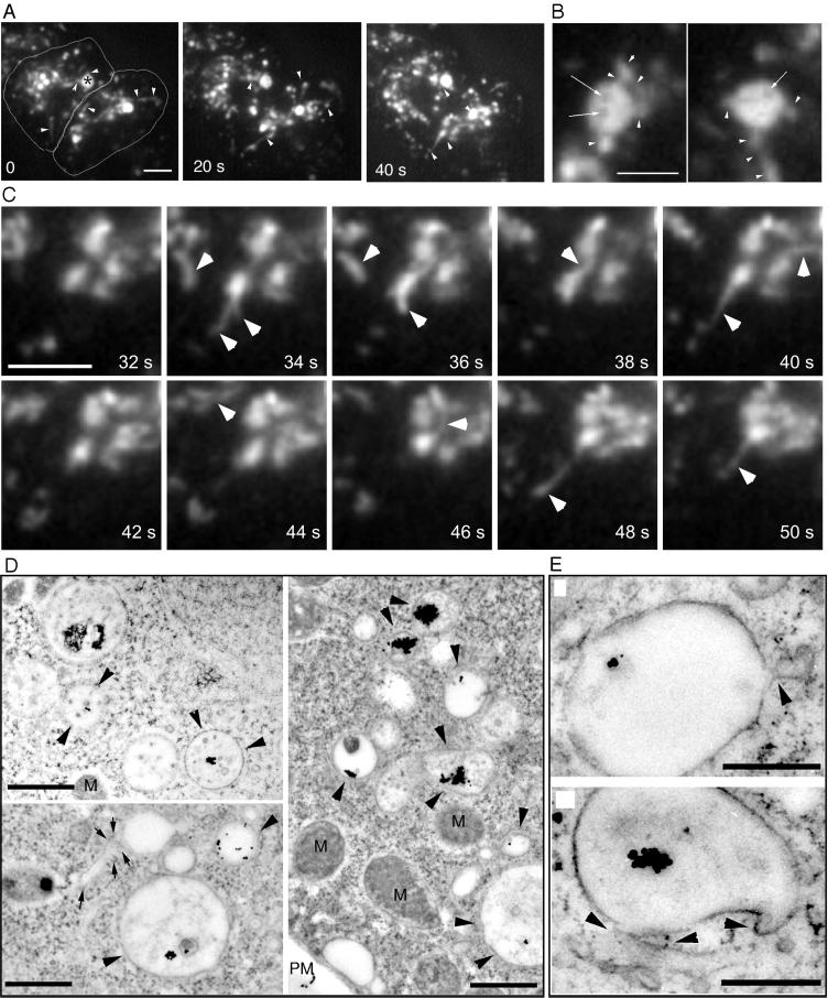
Figure 5.
Tubules and multivesicular bodies. (A) Wild-type cells were fed with rhodamine-green dextran for 30 min and briefly washed with buffer; time-lapse series were recorded with TIRM. The cell boundaries are outlined in the first frame. A very dynamic vacuole with intermediate dextran concentration in the upper left cell (asterisk) repeatedly extended tubular structures (arrowheads); other tubular structures are also marked by arrowheads. The accompanying Movie 4 shows a longer time course (242 s) of the same cell. The movie plays at 6 fps; bar, 10 μm. (B) Selected frames of the vacuole marked by an asterisk in A at a higher magnification, showing again the repeated extension of tubular structures (arrowheads). In addition, the vacuole was inhomogeneously filled with dextran, dark patches in the vacuolar lumen are marked by arrows. Bar, 5 μm. (C) Higher magnification of a vacuole that repeatedly extended tubular structures (arrowheads). Bar, 2 μm. (D) Ultrastructure of endosomes in wild-type D. discoideum cells that ingested BSA-Au particles for 30 min. Some endosomal vacuoles contained internal membranes and aggregated gold particles (arrowheads), a tubular structure is marked by arrows. Bars, 1 μm. (E) Higher magnification of late endosomal vacuoles with extending tubular structures (arrowheads). M, mitochondria. Bars, 0.5 μm.

