Figure 6.
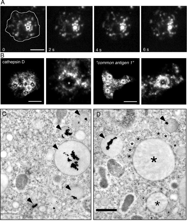
Clusters. (A) Wild-type cells were fed with rhodamine-green dextran for 60 min and briefly washed with buffer; time-lapse series were recorded with TIRM. The cell boundaries are outlined in the first frame. The big central vacuole (asterisk) was surrounded by smaller vesicular structures that continuously moved around and repeatedly contacted this vacuole. The accompanying Movie 5 shows a longer time course (242 s) of the same cell. The movie plays at 6 fps; bar, 10 μm. (B) Immunofluorescence localization of lysosomal enzymes. Antibodies against cathepsin D and against the “common antigen 1,” a mannose-6-sulfate–containing carbohydrate epitope present on D. discoideum lysosomal enzymes such as α-mannosidase and β-glucosidase (Knecht et al., 1984; Freeze et al., 1990), labeled small vesicular structures, that formed “rings of dots” around bigger, unlabeled vacuoles (marked by asterisks and also shown in higher magnification). Bar, 10 μm. (C and D) Ultrastructure of late endosomes in wild-type D. discoideum cells that ingested BSA-Au particles for 60 min. Late endosomes were characterized by electron dense lumen and strongly aggregated gold particles (arrowheads). Some endosomal vacuoles with dark lumen (big asterisk) were surrounded by smaller vesicular structures (small asterisks) reminiscent of the situation in A. Bar, 1 μm.

