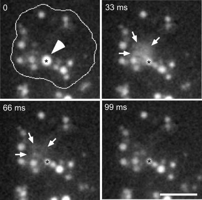
Figure 8.
Exocytosis from the secretory lysosomal compartment. Wild-type cells were fed with rhodamine-green dextran for 2 h and briefly washed with buffer; time-lapse series were recorded with TIRM. The cell boundaries are outlined in the first frame. A big, bright vacuole (asterisk and arrowhead) was almost immobile for some minutes. It suddenly fused with the plasma membrane and egested its content in the extracellular space in the form of a cloud of staining (delimited by arrows). Note that fusion was not complete, because a remnant of the secretory lysosome was still visible after egestion. The accompanying Movie 8 shows a longer time course (1.7 s) of the same cell, it plays at 6 fps; bar, 10 μm.

