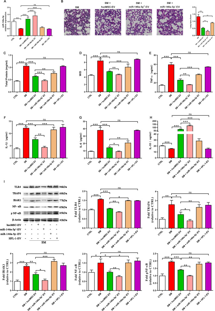
Fig. 6.
MiR-146a-5p delivered by hucMSC-EVs is involved in the hucMSC-EV-mediated therapeutic effects in vivo. ICR mice were subcutaneously injected with diluted SM (30 mg/kg weight). Extracellular vesicles (6 × 10⁸ particles) were injected into the mice through the tail vein on the first and third days after SM exposure. On the fifth day, the lung tissues were collected for subsequent experiments. A miR-146a-5p expression in mouse lung tissues was examined by qRT–PCR (n = 6 mice/group). B Representative histological micrograph analysis (× 200, scale bar = 100 μm) and histopathological scores (n = 3 mice/group). C, D Comparison of BALF protein and the wet/dry weight ratio in SM-exposed mice (n = 6 mice/group). E–H ELISA showing the expression of the inflammatory factors TNF-α, IL-1β, IL-6, and IL-10 in mouse lung tissues (n = 6 mice/group). I The expression of TLR4/NF-κB signaling pathway-related proteins isolated from lung tissues was visualized by western blotting (n = 3 mice/group). Full-length blots are presented in Additional file 1: Fig. S7. The data are presented as the mean ± SD of individuals included in each group and representative of at least three independent experiments. *P < 0.05; **P < 0.01; and ***P < 0.001

