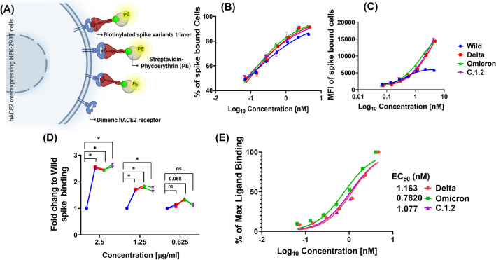
Figure 4.
Potency of spike variants affinity to dimeric hACE2 receptors present on surface of the cells. Schematic representation of in vitro flow cytometry binding assay to estimate binding affinity of spike variants (A). The 293T-hACE2 cells were surface stained with biotinylated trimeric spike variants at different concentration. Percentage (B) and level (MFI) (C) of spike trimer bound to cells was quantified using streptavidin-PE conjugate in flow cytometry. The fold change of Delta, Omicron and C.1.2 binding (MFI) related to Wild was calculated at indicated concentration (D). Percentage of maximum spike binding was calculated from MFI, plotted against concentration and a non-linear curve fitting was used to estimate the EC50 of spike variants (E).

