
Figure 2. Intracranial stimulation of the human amygdala induces apnea.
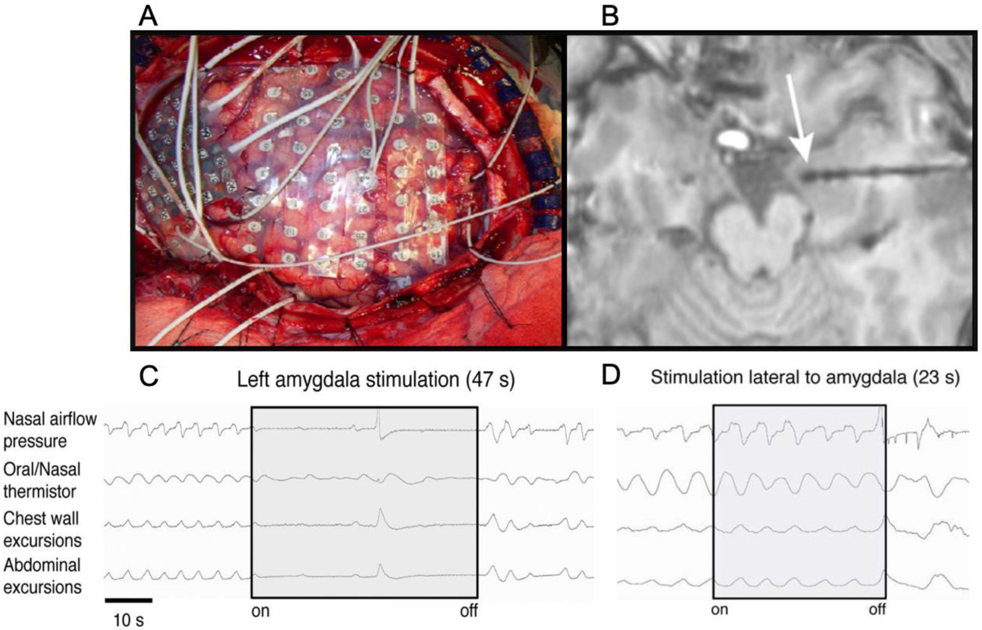
(images from Dlouhy et al., 2015). (A) For many years, neurosurgeons have applied large arrays of electrodes inside the brain of epilepsy patients to identify the source of their seizures prior to resecting the epileptogenic tissue. These intracranial electrodes were mainly used for passively recording neuronal activity. More recently, neurosurgeons have started using the electrodes to stimulate the underlying tissue by applying extended trains of electrical pulses at 50 Hz while simultaneously recording changes in respiration. (B) Depth electrodes stimulated in the vicinity of the amygdala (white arrow) reliably trigger apnea (C), whereas stimulation of electrodes outside of the amygdala does not disrupt respiration (D).