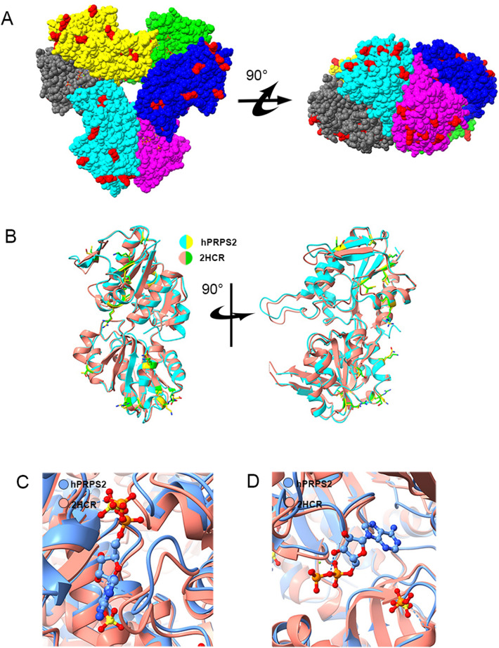
Fig. 4.
Structural comparison of hPRPS2 and hPRPS1 (2HCR). A The difference of amino acids between hPRPS1 (2HCR) and hPRPS2. There are 15 amino acid residues differences between human PRPS1 monomer and human PRPS2 monomer. Most of the different amino acids are located on the surface of the hexamer. The amino acid residues with differences are marked in red. B Comparison of hPRPS1 and hPRPS2 monomers. The monomer of human PRPS1 is in red and human PRPS2 is in cyan. The amino acid residues with difference in human PRPS1 and human PRPS2 are green and yellow, respectively. C Comparison of allosteric site and RF loop. There is an ADP at the allosteric site of human PRPS2, while SO42− in human PRPS1 can bind to allosteric site and another site. The chain of human PRPS1 (2HCR) is in red and of human PRPS2 is in blue. D Comparison of human PRPS1 (2HCR) and human PRPS2 active sites. In human PRPS2, ADP and magnesium occupy the ATP binding site in the active site, which is empty in the ATP binding site of human PRPS1 (2HCR). SO42− and PO43− are found in the R5P binding sites of human PRPS1 (2HCR) and human PRPS2, respectively

