
Figure 1: Ex vivo electroretinogram specimen holder and recording setup.
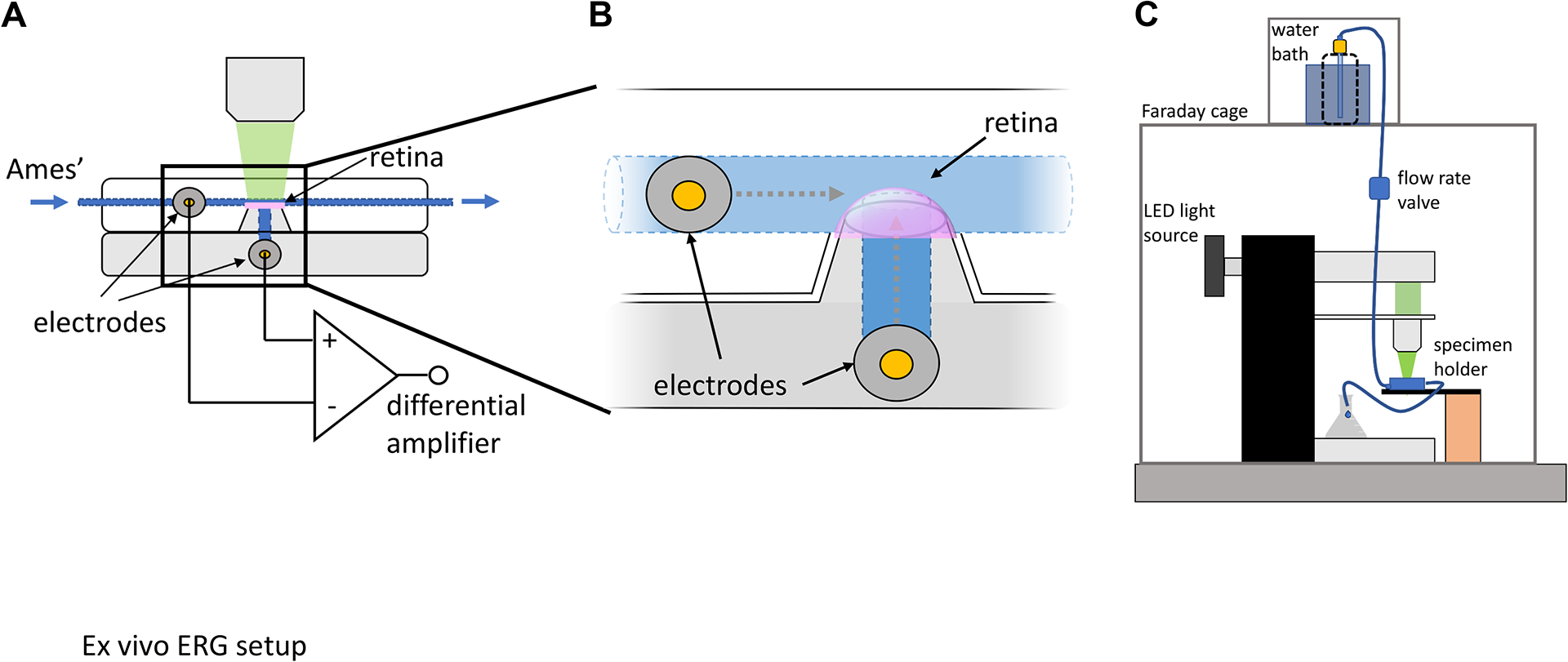
(A, B) The ex vivo ERG specimen holder comprises a dome to mount the isolated retina, which is connected to a perfusion line to continuously deliver Ames’ medium. Electrodes are connected through narrow channels to both the photoreceptor side of the retina via the perfusion line and the inner retina through the filter paper glued to the dome. These electrodes are connected to a differential amplifier, which enables measurement of potential differences in the retina in response to light stimuli. (C) The specimen holder is placed onto the stage of an inverted microscope, which has been modified to deliver light flashes and connected to the perfusion line, which delivers heated, oxygenated Ames’ medium by gravity. The entire recording setup is shielded by a Faraday cage to minimize electrical noise. This figure has been modified from9. Abbreviation: ERG = electroretinogram.