. 2023 May 27;15(1):2217964. doi: 10.1080/19420862.2023.2217964

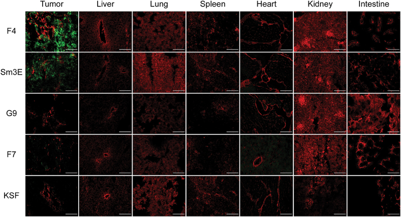
Figure 3.

Immunofluorescence staining of the tumor, liver, lung, spleen, heart, kidney, and intestine from the groups F4, Sm3E, G9, F7, and KSF. Strong green staining is present in the tumor of the F4 group and at a slightly lower intensity in the Sm3E group. Minimal green staining is present in the tumor of the F7 group. No green staining is present in the tumors of the G9 and KSF groups and in all healthy organs.

Ex vivo immunofluorescence-based biodistribution analysis. Immunofluorescence analysis assessed tumor targeting of new anti-CEA antibodies in IgG format. Two hundred micrograms of IgG-FITC were injected intravenously into LS174T-bearing mice. Tumors were excised 24 hours after injection. IgG-FITC was detected in green; blood vessels were detected through CD31 staining (red). 20× magnification, scale bars = 100 μm.