
FIGURE 2.
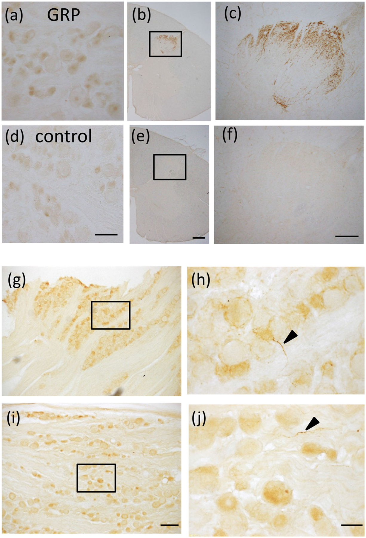
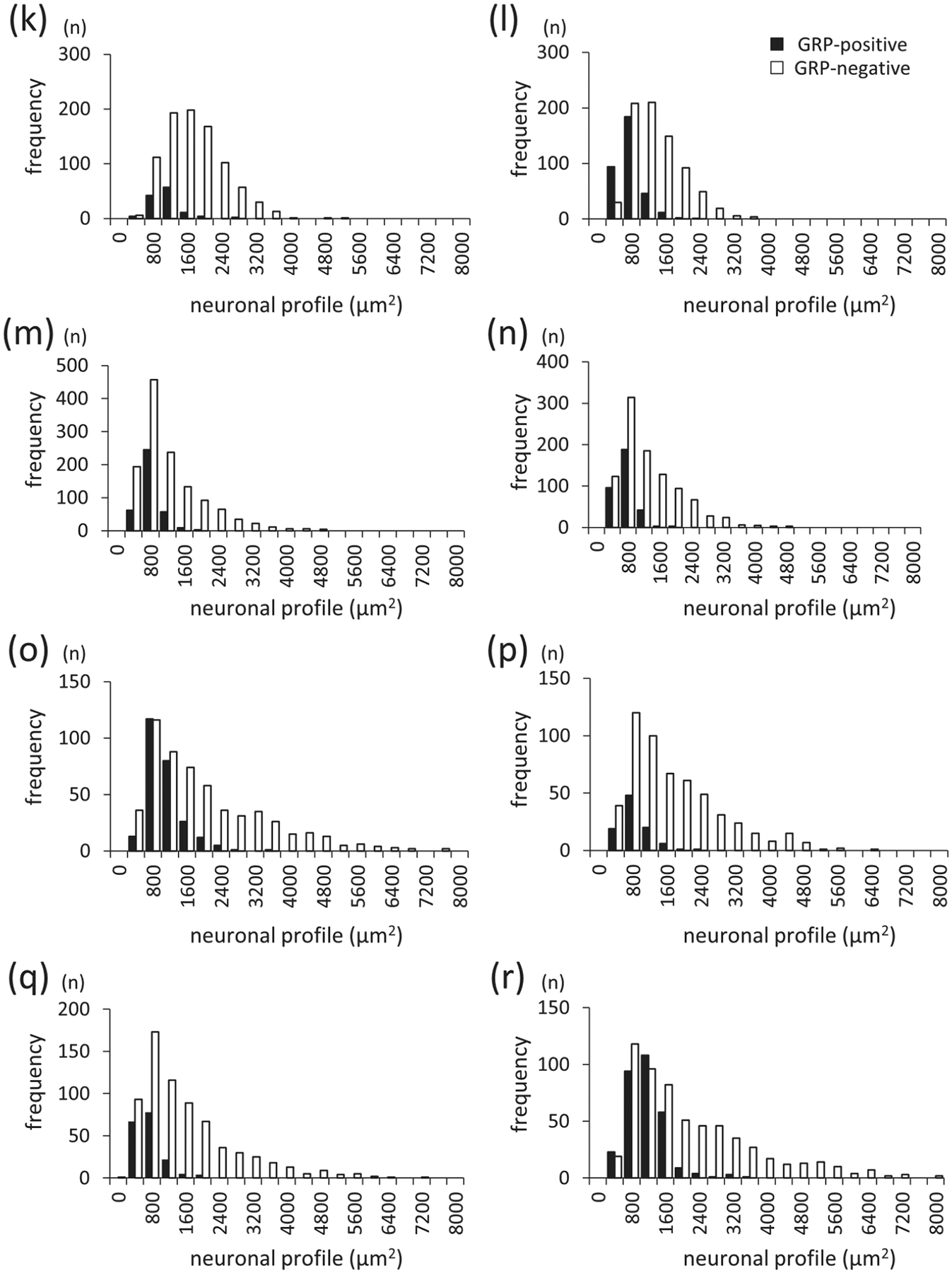
GRP expression and distribution in the trigeminal ganglion (TG) and dorsal root ganglion (DRG) in macaque monkeys, (a–f) Immunohistochemical staining using GRP antiserum in the DRG and cervical spinal cord in adult macaque monkeys. GRP-immunoreactivity (ir) was observed in some neurons in the macaque monkey DRG (a) and in dense fiber projections to the superficial layers of the cervical spinal cord (b, c). Controls in which anti-GRP antiserum was preabsorbed with an excess of antigen peptide (50 μg/mL) showed an absence of GRP expression in the DRG (d) and dorsal horn of the spinal cord (e, f). (c and f) are enlargements of the boxed areas in (b and e), respectively. Bars = 100 μm (d); 500 μm (e); 200 μm (f). (g–j) GRP expression in theTG and cervical DRG of adult male macaque monkeys. GRP-ir neurons were mainly observed in the small-sized TG neurons (g, h) and DRG neurons (i, j). (h) and (j) are enlargements of the boxed areas in (g) and (i), respectively. Bars = 200 μm (i); 50 μm (j). (k–r) Histograms of size distribution of GRP-positive and GRP-negative neurons in the monkey TG (k–n) and DRG (o–r). GRP-ir neurons were predominantly the small- and medium-sized TG and DRG neurons. GRP-positive TG neurons were 12% in the 8.3 kg female (k), 31% in the 7.2 kg female (I), 23% in the 2.7 kg male (m), and 25% in the 2.9 kg male monkey (n). GRP-positive DRG neurons were 31% in the 8.3 kg female (o), 15% in the 7.2 kg female (p), 20% in the 2.7 kg male (q), and 33% in the 2.9 kg male monkey (r). The average of GRP-positive TG neurons was 23% and DRG neurons was 25% in the four monkeys