
FIGURE 4.
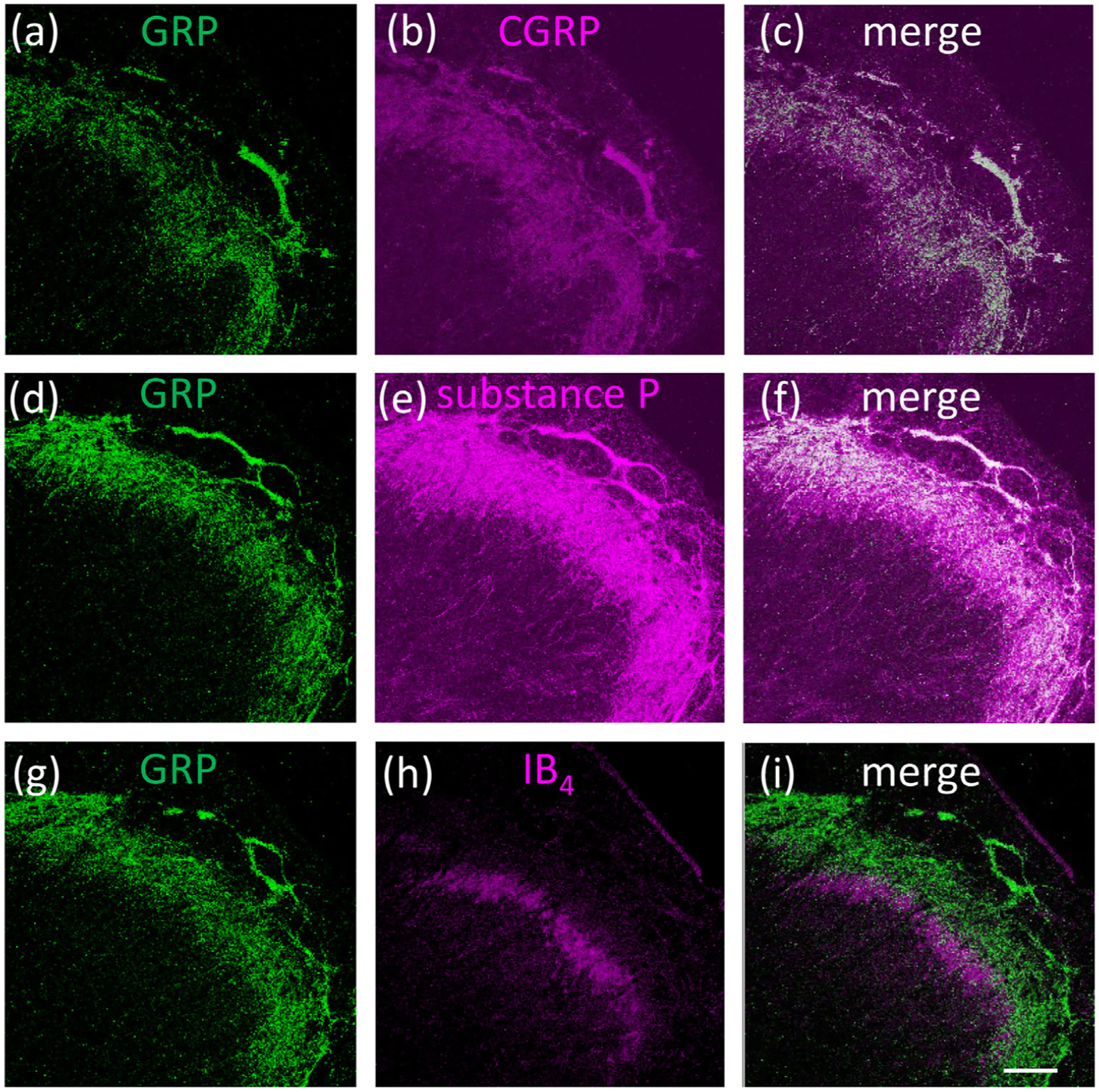
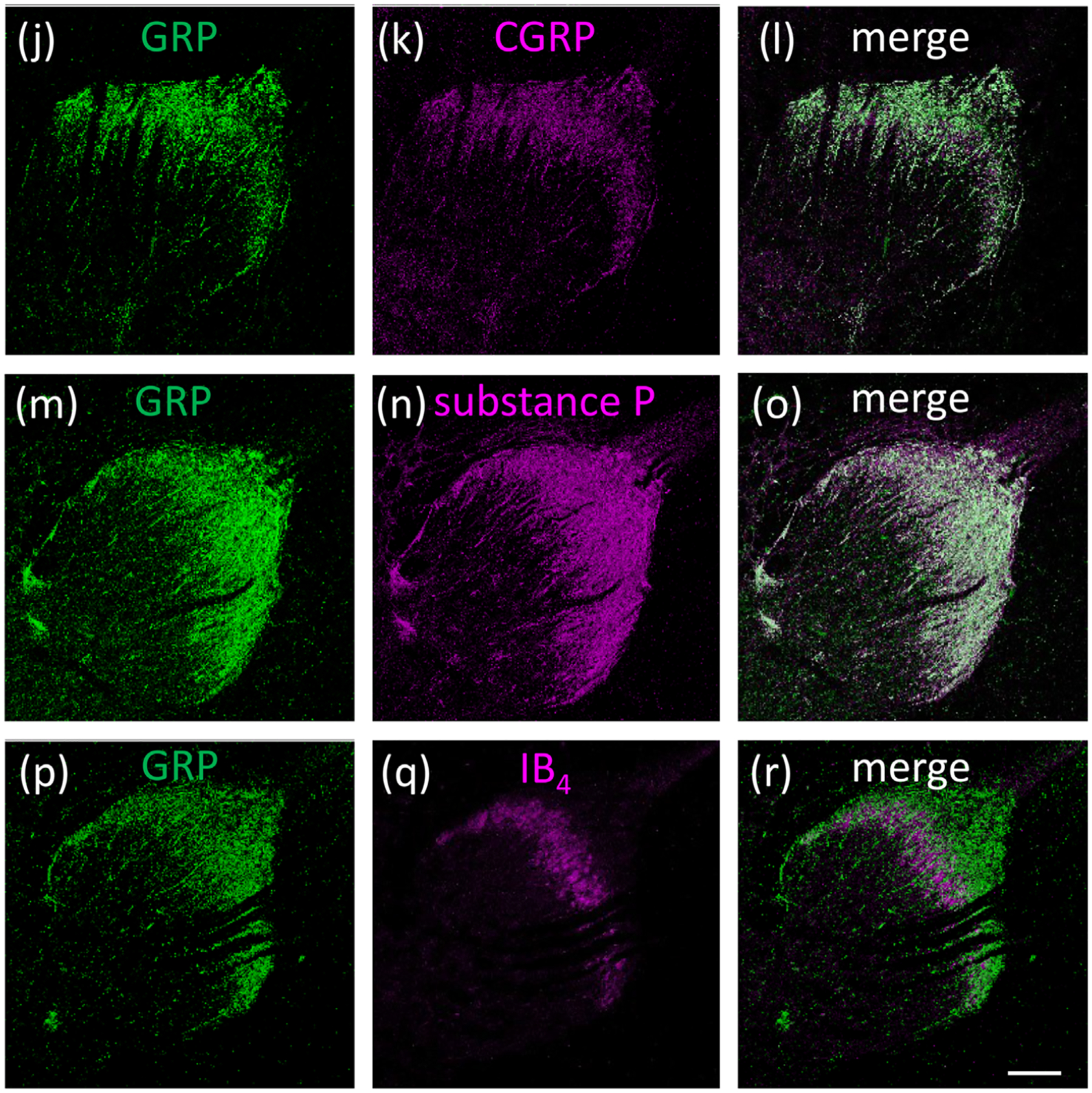
Double fluorescence of the GRP (green) and superficial layers markers (magenta) of Sp5C and cervical spinal dorsal horn in adult macaque monkeys. Many GRP-ir fibers were colocalized with the marker for peptidergic neurons CGRP (a–c) and substance P (d–f) in the Sp5C. GRP-ir fibers were not colocalized with the marker for nonpeptidergic neurons IB4 in the Sp5C (g–i). Most GRP-ir fibers were colocalized with CGRP(j–l) and substance P (m–o) in the superficial layers of the cervical spinal cord. GRP-ir fibers were not colocalized with IB4 (p–r) in the cervical spinal dorsal horn. Bars = 200 μm (i, r)