
FIGURE 6.
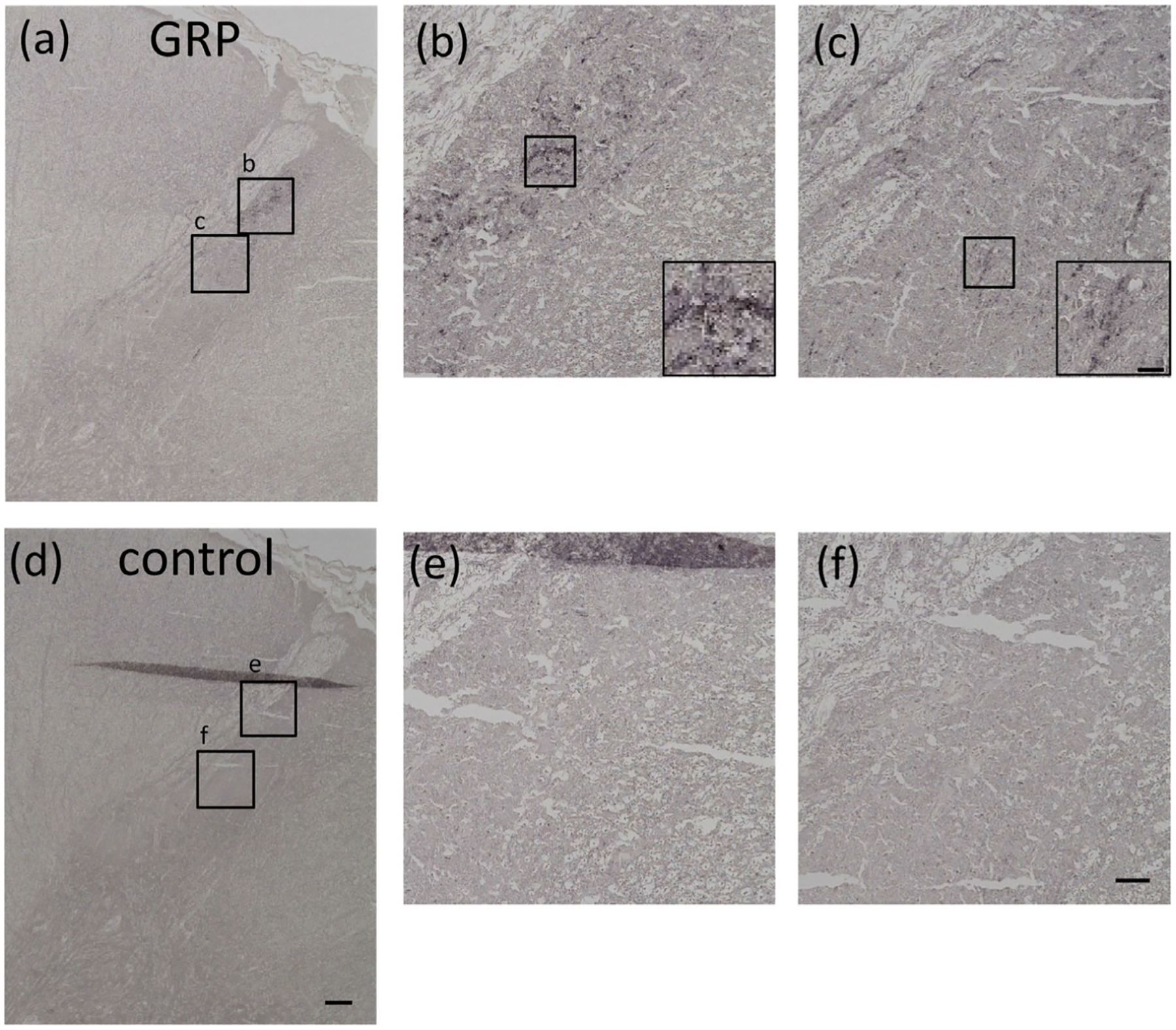
Immunohistochemical staining using GRP antiserum in the spinal cord in adult human male. GRP-ir was observed in dense fiber projections to the superficial layers of the transition area between thoracic and lumbar spinal cord (a–c). Controls in which anti-GRP antiserum was preabsorbed with an excess of antigen peptide (50 μg/mL) showed an absence of GRP expression in the spinal dorsal horn (d–f). (b, c, e, and f) are enlargements of the boxed areas in (a) and (d), respectively. Bars = 200 μm (d); 50 μm (f); 10 μm (inserted boxed area of c)