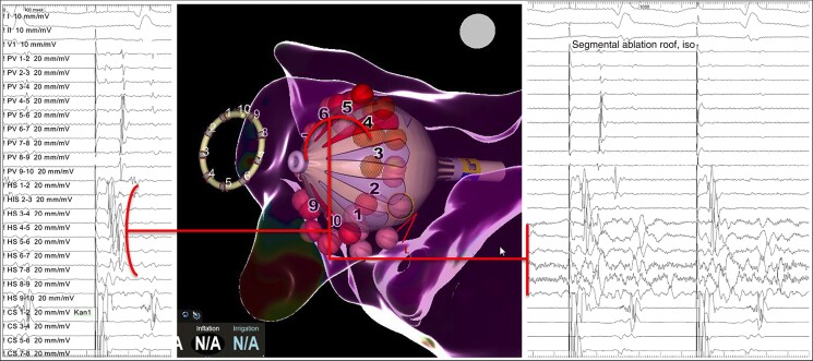
Figure 2.
Example of a segmental isolation of a left superior pulmonary vein. Notice the earliest activation of the PV in the Heliostar (HS) electrodes 4 to 7 (left panel), preceding the signals collected distally by the Lasso-star (PV). In the right panel, real time PVI could be recorded during localized application of RFC at these electrodes. PV, pulmonary vein; PVI, pulmonary vein isolation; RFC, radiofrequency current.

