
Figure 4.
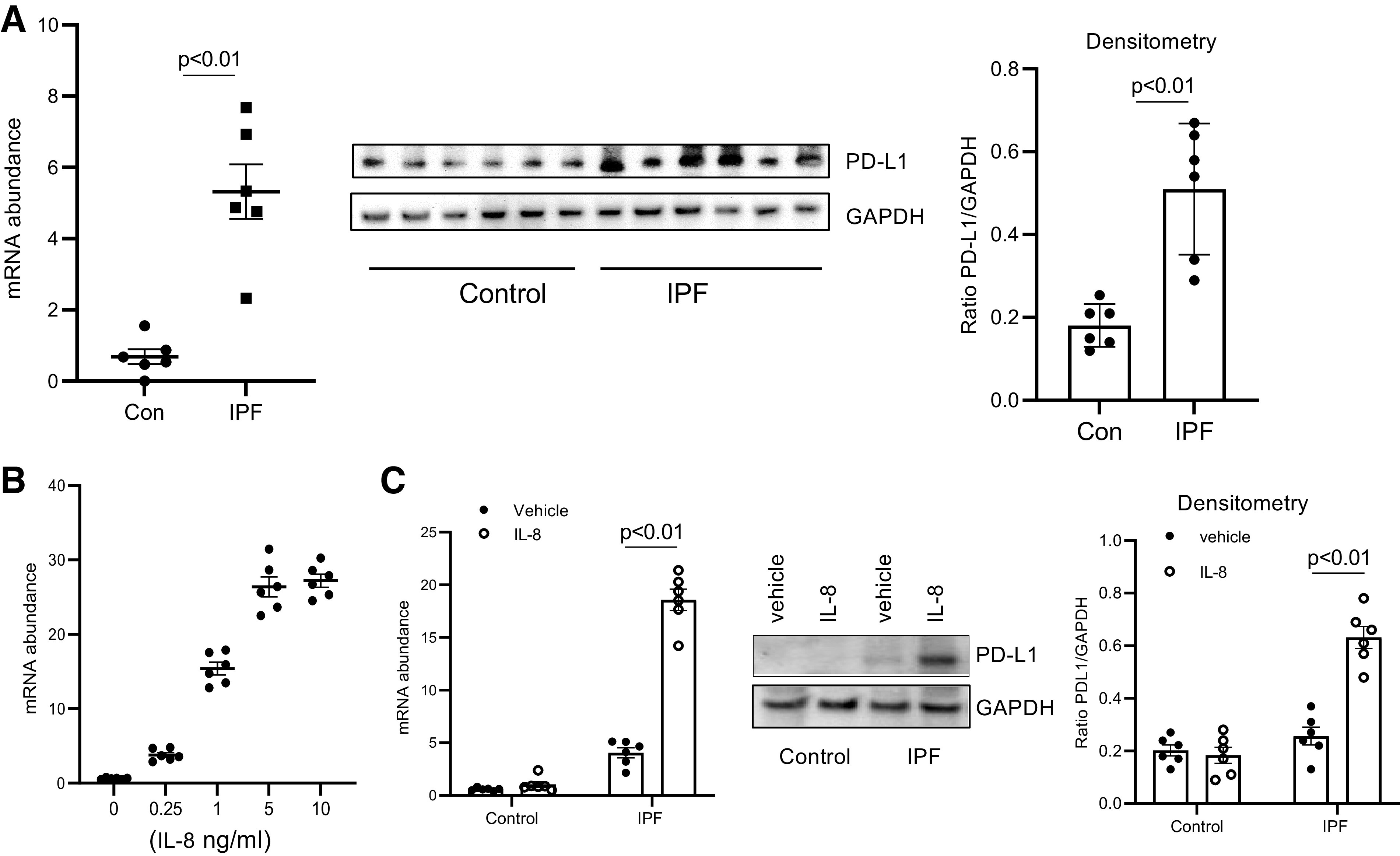
Programmed death ligand 1 (PD-L1) levels are elevated in idiopathic pulmonary fibrosis (IPF) mesenchymal progenitor cells (MPCs). A: PD-L1 expression was quantified in IPF and control (Con) MPCs (n = 6 IPF and control MPC cell lines each) by quantitative PCR (Q-PCR) (left) and Western blot analysis (center). GAPDH = loading control. Densitometry values summarizing Western blot data are on right. B: IPF MPCs were treated with various amounts of IL-8. PD-L1 levels were quantified by Q-PCR. C: IPF and control MPCs (n = 6 cell lines each) were treated with IL-8 (5 ng) or vehicle control. PD-L1 levels were quantified by Q-PCR (left) and Western blot analysis (shown is a respresentative Western blot quantifying PD-L1 levels in 1 IPF and 1 control MPC cell line; center). GAPDH = loading control. Densitometry values summarizing Western blot data are on right.