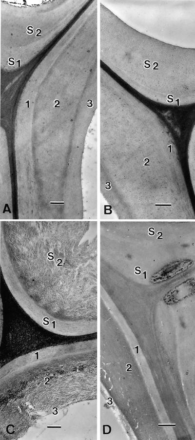
Figure 5.
Ultrastructural morphology of fibers and vessels observed in TEM after transverse section of the stem. A through C, periodic acid-thiocarbohydrazide-Ag proteinate staining. D, Uranyl acetate staining. A, Wild-type plant: the fiber secondary wall and its sublayers S1 and S2 appear very compact. Note the typical subdivision of the vessel wall in the three sub-layers noted 1, 2, and 3. B, B10 (ASCOMT I) single transformant. No visible ultrastructural alteration. C, B31 (ASCCR) single transformant exhibits a pronounced loosening of its cellulosic framework in S2 of the fiber walls. Sub-layers 2 and 3 of the vessel wall also are affected. D, Double transformant from B31 × B10 cross: Only a slight alteration of ultrastucture is detected in sub-layer 3 of vessel; no particular loosening in sub-layer 2 of the vessel wall is visible (the white cracks in internal S2 are artifacts due to embedding). Bars in A and C represent 0. 5 μm; in B and D, they represent 0.7 μm.

