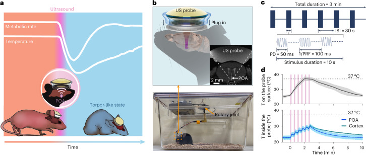
Fig. 1. Ultrasound device for inducing a torpor-like hypothermic and hypometabolic state.
a, Illustration of ultrasound (US)-induced torpor-like state. b, Illustration of the wearable US probe (top). The probe was plugged into to a baseplate that was glued on the mouse’s head. MRI of the mouse head with the wearable US probe shows that ultrasound was noninvasively targeted at the POA (insert). Photograph of a freely moving mouse with the wearable US probe attached is shown at the bottom. c, Illustration of the US stimulation waveform used in this study. ISI, inter-stimulus interval; PD, pulse duration; PRF, pulse repetition frequency. d, Calibration of the temperature (T) rise on the surface (top) and inside (bottom) the US probe. The temperature inside the probe was measured between the piezoelectric material and the mouse head when US probes were targeted at the POA or the cortex. Cortex was selected as an off-target control. US sonication is indicated by the pink bars. n = 6 mice (top) and n = 4 mice (bottom). Solid lines and shadows denote the mean ± s.e.m.

