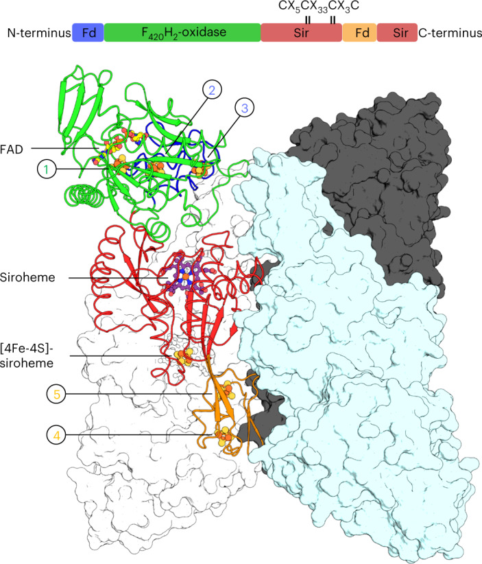
Fig. 1. Domain and structural organization of MtFsr.

Visualization of MtFsr domains (top panel). The [4Fe‒4S] cluster-binding motif in the proximity of the siroheme is highlighted. The main panel shows the tetrameric arrangement of MtFsr. Three chains are represented in the surface and colored in white, black and cyan. One monomer of MtFsr is represented as a cartoon and colored according to the top panel. [4Fe‒4S] clusters are numbered on the basis of their position in the electron relay going from the FAD to the siroheme. The siroheme, FAD and the [4Fe‒4S] clusters are represented by balls and sticks. Carbon, nitrogen, oxygen, sulfur and iron atoms are colored as purple (siroheme)/light yellow (FAD), blue, red, yellow and orange, respectively. Fd and Sir stand for ferredoxin domain and sulfite reductase domain, respectively.
