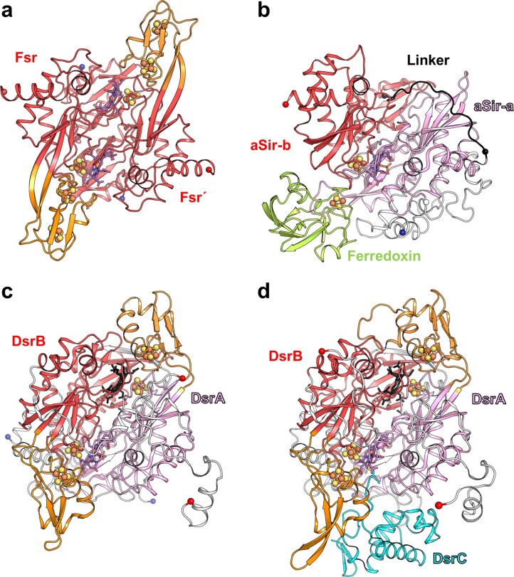
Extended Data Fig. 7. Overall structural comparison between Fsr, aSir and dSir.
Cut-through view shown in cartoon of one dimer for Fsr and DsrAB. Ligands are shown as balls and sticks. a, The sulfite reductase domain with the inserted ferredoxin domain of MtFsr. Fsr´ corresponds to the opposite monomer. b, aSir from Zea mays and its [2Fe-2S]-ferredoxin coloured in light green (PDB 5H92). c, DsrAB from A. fulgidus (PDB 3MM5) and d, DsrABC from D. vulgaris (PDB 2V4J). The inserted ferredoxin domains of Fsr, DsrA and DsrB are coloured in orange. The catalytic siroheme in DsrAB is coloured in purple and the structural siroheme is coloured in black. DsrAB from D. vulgaris contains sirohydrochlorin instead of siroheme.

