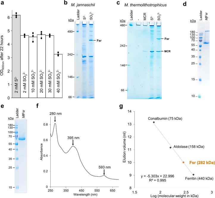
Extended Data Fig. 2. Physiological and biochemical profiles of Fsr from Methanococcales.
a, Final OD600 nm of M. thermolithotrophicus grown on sulfide (S2-) and different sulfite (SO32−) concentrations as a sole sulfur source after 22 hours (mean ± s.d., n = 3 biologically independent replicates). b, c, hrCN-PAGE of cell extracts (12 µg loaded) from M. jannaschii (b, n = 1 independent experiment) and M. thermolithotrophicus (c, n = 3 independent experiments), grown on 2 mM Na2S or 2 mM Na2SO3 as a sole sulfur source. Purified MCR from M. thermolithotrophicus (1.7 µg loaded) was used as a control for the hrCN-PAGE54. d, e, SDS-PAGE profile of purified MjFsr (d, n = 1 independent experiment) and MtFsr (e, n = 3 independent experiments). f,. UV-visible spectrum of 0.33 mg MtFsr measured anaerobically (100 % N2) in 25 mM Tris-HCl, pH 7.6, 150 mM NaCl, 10 % v/v glycerol and 2 mM DTT. MtFsr displays the typical spectra of [Fe-S]-cluster and siroheme containing enzymes55, similar to the UV spectrum of MjFsr previously determined exhibiting three peaks at 280 nm, 395 nm and 593 nm5. g, Molecular weight estimation of MtFsr via size exclusion chromatography (Superdex 200 Increase 10/300 GL from GE Healthcare). Apparent molecular weight of purified MtFsr (monomeric molecular weight = 69.145 kDa) was estimated to 282 kDa. MtFsr is therefore apparently organized as a homotetramer (theoretical molecular weight of the protein in the homotetramer: 276.58 kDa).

